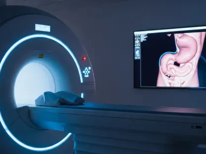

The landscape of modern oncology is shifting toward a highly personalized model where diagnostic imaging and therapeutic intervention are no longer separate silos but two sides of the same molecular coin. Theranostics, a sophisticated medical strategy, utilizes a single targeting molecule that can be labeled with either a diagnostic radioisotope for initial detection or a therapeutic one for targeted destruction of cancer cells. This integrated methodology allows clinicians to “see what they treat and treat what they see,” ensuring that the radioactive drug is reaching its intended destination before the full therapeutic course begins. By leveraging the specific biological signatures of tumor cells, this approach offers a level of precision that traditional systemic chemotherapy or external-beam radiation often struggles to match, marking a significant departure from one-size-fits-all medical protocols.
The clinical validation of radiopharmaceutical therapy (RPT) has gained significant momentum with the regulatory approval and successful implementation of drugs such as Lutathera and Pluvicto. Lutathera has transformed the management of neuroendocrine tumors by targeting somatostatin receptors, while Pluvicto has become a vital tool in treating metastatic castration-resistant prostate cancer by focusing on the prostate-specific membrane antigen. These drugs function by binding a tumor-seeking ligand to a potent isotope like Lutetium-177, which emits localized beta radiation to kill malignant tissue while sparing the surrounding healthy organs. Currently, over 70 companies are aggressively pursuing the expansion of this technology, with numerous candidates in phase-three trials aimed at treating breast cancer, lung cancer, and lymphoma at much earlier stages of disease progression than previously possible.
Challenges in Biological Variability and Standardization
Addressing Technical and Biological Inconsistencies
A primary challenge currently hindering the universal adoption of radiopharmaceutical therapy is the inherent lack of standardized treatment protocols and robust quality assurance methodologies across different healthcare systems. In the realm of traditional external-beam radiotherapy, medical physicists maintain absolute control over the radiation source, allowing for high-precision beam steering and predictable energy deposition within the patient’s body. Conversely, the delivery of a dose in RPT is governed by a complex and often unpredictable interplay of biological and biochemical factors, including individual blood flow patterns, varying receptor densities on tumor surfaces, and the patient’s unique metabolic clearance rates. These variables introduce a layer of uncertainty that makes it difficult to predict exactly how much radiation will be absorbed by a specific tumor or a critical organ like the kidney.
This biological variability frequently manifests as significant inconsistencies in dosimetry results—the measurement and calculation of the radiation dose absorbed by human tissue. Recent investigations have shown that even highly trained medical physicists can produce dosimetry calculations that fluctuate by as much as 30% when analyzing the exact same patient data due to differences in software algorithms and manual interpretation. Without a unified framework to harmonize these measurements, the medical community finds it difficult to establish universal radiation dose-effect curves that are essential for defining safe and effective treatment limits. This lack of standardization not only complicates clinical decision-making but also poses a potential risk to patient safety, as clinicians struggle to find the optimal “dose-window” that maximizes the destruction of malignant cells without causing irreversible toxicity to healthy bone marrow or renal tissues.
The Impact of Variable Software and Expert Interpretation
The diversity of software packages and mathematical models used to calculate absorbed doses further exacerbates the problem of inconsistency in clinical settings. Currently, different clinics may utilize various proprietary or open-source tools to process SPECT/CT imaging data, leading to a situation where a patient might receive a different dose recommendation depending on which hospital they visit. This variability is often compounded by the level of experience of the local physics team, as the manual contouring of organs and the selection of background subtraction methods vary significantly between operators. Such discrepancies make it nearly impossible to conduct large-scale, multi-center clinical trials with the level of data integrity required to set new global standards for cancer care.
To bridge this gap, there is an urgent need to transition from the current fixed-activity models—where every patient receives the same amount of a drug—to a more rigorous, physics-based approach that accounts for individual biochemical differences. By standardizing the way that imaging data is captured and processed, the industry can ensure that the “activity” of a radioactive drug translates into a predictable “absorbed dose” in the tissue. Establishing these rigorous standards is the only way to move RPT from a last-resort treatment for metastatic patients to a primary, front-line therapy. This shift requires not just better technology, but a fundamental change in the collaborative relationship between nuclear medicine and radiation oncology departments, ensuring that the same level of physical rigor applied to linear accelerators is applied to radioactive injections.
Global Initiatives for Uniformity
Implementing the Precision Dosimetry Imaging Biomarker Project
The Precision Dosimetry Imaging Biomarker (PDIB) project represents a comprehensive international effort designed to bring the exacting standards of radiation physics into the rapidly evolving nuclear medicine environment. This ambitious initiative is built upon three critical pillars: the establishment of secondary standards calibration laboratories, the harmonization of SPECT/CT scanner calibrations, and the standardization of dosimetry calculation workflows. By creating a global network of specialized laboratories, such as those at the University of Iowa and the Belgian Nuclear Research Centre, the project ensures that the radionuclide calibrators used in local clinics are tuned with an activity uncertainty of less than 3%. This foundational accuracy is essential for ensuring that when a physician prescribes a specific amount of radioactivity, the patient receives exactly that amount, regardless of the geographic location of the treatment center.
Furthermore, the PDIB project focuses on the harmonization of quantitative imaging data to ensure that a scan taken in one hospital provides the same diagnostic information as a scan taken in another. By utilizing standardized physical phantoms and samples verified by national metrology institutes like NIST, the project enables clinicians to calibrate SPECT/CT scanners across various manufacturers and models. This uniformity allows for the development of standard operating procedures for software and mathematical models, which are tested against curated datasets to minimize inter-user variability. As a result, the project ensures that a patient’s treatment plan remains consistent and reproducible, regardless of the facility or the specific software package utilized for the final dose calculation, providing a reliable basis for personalized cancer therapy on a global scale.
Advancing Quantitative Imaging and Phantom Validation
The role of physical phantoms in the PDIB project cannot be overstated, as they provide the necessary ground truth for calibrating complex imaging equipment. These phantoms are essentially physical models that mimic human anatomy and are filled with known quantities of radioactive isotopes to test how accurately a scanner can detect and quantify radiation. By shipping these standardized tools to imaging sites worldwide, the project can identify and correct variations in scanner performance caused by aging components or different reconstruction algorithms. This level of technical scrutiny ensures that the visual “brightness” of a tumor on a PET or SPECT scan is a true reflection of the radioactive concentration, which is the most critical data point needed for calculating the subsequent therapeutic dose.
In addition to hardware calibration, the PDIB initiative is driving the development of specialized “digital phantoms” and curated datasets that allow software developers to benchmark their dosimetry tools against a gold standard. By analyzing 177Lu-DOTATOC data for kidneys and tumors through these standardized digital environments, the medical community can identify which calculation methods are most resistant to human error or algorithmic bias. This dual approach of physical and digital validation creates a robust ecosystem where every step of the theranostic workflow—from the initial injection to the final dose report—is cross-referenced against a reliable standard. This systematic reduction of technical noise allows the true biological response of the patient to become the primary focus of the treatment, paving the way for more effective and safer oncological interventions.
The Future of Personalized Clinical Practice
Dosimetry-Modulated Therapy and Professional Integration
The ultimate objective of these global standardization efforts is the widespread implementation of dosimetry-modulated therapy, a dynamic approach that moves beyond static dosing schedules. Rather than receiving a pre-determined, fixed amount of radioactive activity over several cycles, patients can benefit from a personalized regimen where the treatment is adjusted based on real-time serial imaging. For example, if a SPECT/CT scan after the first cycle of a drug like Pluvicto reveals a rapid and significant tumor response, the physician might choose to conclude the treatment early, thereby preventing unnecessary radiation exposure and reducing the risk of side effects. Conversely, if the scan shows that the tumor is resistant but the radiation levels in healthy organs remain well below safety thresholds, the clinician can safely increase the injected activity for the next cycle to enhance the curative potential.
This evolution in clinical practice is simultaneously reshaping the professional landscape by merging the biological insights of nuclear medicine with the technical and quantitative rigor of radiation oncology. As the market for nuclear medicine continues to expand exponentially, there is a critical need for radiation oncology physicists to bring their expertise in complex radiation delivery and safety protocols into the theranostic fold. This multidisciplinary collaboration is effectively breaking down traditional departmental silos, creating a unified medical department centered on the most effective use of radiation. By integrating the skills of both specialties, healthcare institutions can provide a more holistic approach to cancer care, ensuring that the precision of molecular targeting is matched by the precision of physical measurement and safety monitoring.
Bridging the Gap Between Research and Bedside Application
As these professional roles merge, the focus is shifting toward the creation of integrated theranostic centers where the workflow is optimized for rapid data sharing between diagnostic and therapeutic teams. In these environments, the medical physicist acts as the bridge, ensuring that the quantitative data from a diagnostic scan is immediately and accurately translated into a treatment plan. This requires not only high-level technical skills but also a deep understanding of the biological behavior of radiopharmaceuticals. The transition toward this integrated model is being accelerated by new training programs and certifications that encourage radiation oncology physicists to specialize in molecular radiotherapy, addressing the current shortage of experts in this high-growth field.
Moreover, this professional integration is fostering a new culture of evidence-based practice where every treatment decision is backed by standardized data. The move away from empirical “one-size-fits-all” dosing toward individualized, physics-led therapy is expected to significantly improve patient outcomes and survival rates. By treating the patient’s body as a dynamic system that changes throughout the course of therapy, clinicians can adapt their strategies in real-time, offering a level of flexibility that was previously impossible. This paradigm shift ensures that the medical community is not just administering drugs, but is actively managing the radiation environment within each patient to achieve the best possible therapeutic index, marking a new era of sophistication in precision oncology.
Technological Infrastructure and Industrial Support
Industry leaders are playing a vital role in supporting this clinical transition by providing the specialized hardware and software infrastructure necessary for large-scale theranostic applications. Modern software platforms, such as the ec² platform, now offer comprehensive digital traceability for the entire radiopharmaceutical lifecycle, from initial production in the radiopharmacy to final administration at the bedside. These systems provide a robust digital trail that ensures regulatory compliance and significantly reduces the risk of human error in an environment where every dose is custom-made for a specific patient. By automating the tracking of radioactive decay and patient-specific metrics, these tools allow busy clinics to scale their theranostic programs without compromising the safety or accuracy of the treatment.
On the physical side of the infrastructure, high-precision instruments such as Capintec dose calibrators and specialized SPECT calibration phantoms are providing the foundation for the rigorous quality assurance demanded by the PDIB project. These tools enable clinics to perform the absolute activity measurements required to maintain the tight uncertainties necessary for personalized dosimetry. When integrated with advanced radiation shielding and real-time monitoring systems, these technological suites create a safe and efficient environment for both patients and staff. By bridging the gap between experimental research and high-volume clinical care, these industrial solutions are providing the essential tools needed to handle potent radioactive drugs with the same level of confidence and traceability as traditional medical interventions.
Scaling Precision Care Through Integrated Solutions
The integration of these technological suites allows healthcare institutions to effectively transition their theranostic programs from small-scale, experimental niches to standardized, high-volume patient care centers. As the complexity of radioactive drugs increases—particularly with the rise of alpha-emitters which require even more precise handling—the role of automated, high-precision hardware becomes even more critical. These systems act as a safety net, ensuring that even as the volume of patients increases, the quality of care remains consistently high. By providing a unified platform for measurement, tracking, and safety, industrial partners are enabling hospitals to treat more patients with greater confidence, ultimately making these life-saving therapies more accessible to the general population.
Furthermore, the data generated by these integrated systems is becoming a valuable resource for continuous improvement in clinical protocols. By aggregating anonymized data from thousands of treatments, researchers can identify patterns and refine the mathematical models used for dosimetry, leading to even more accurate treatment planning in the future. This feedback loop between the clinic and the industry ensures that the technology is constantly evolving to meet the real-world challenges faced by oncologists. As these systems mature and become more interconnected, they will provide the essential data backbone for a truly global standard of precision cancer care, where every microcurie of radiation is accounted for and optimized according to the unique biological profile of every patient.
Strategic Implementation for Healthcare Facilities
To capitalize on the advancements in theranostics, healthcare administrators should prioritize the modernization of their nuclear medicine infrastructure, ensuring that hardware and software are compatible with the latest standardization protocols like those from the PDIB project. Facilities must invest in specialized training for their medical physics teams to bridge the expertise gap between radiation oncology and molecular imaging, fostering a collaborative environment that can handle the complexities of dosimetry-modulated therapy. Furthermore, adopting integrated digital platforms for dose tracking and regulatory compliance will be essential for scaling these programs safely as patient demand increases. By establishing robust quality assurance frameworks now, institutions will be well-positioned to integrate the next generation of alpha-emitting radiopharmaceuticals into their primary oncological workflows. The transition toward standardized theranostics is not merely a technical upgrade but a strategic move toward a more effective, data-driven model of cancer care that promises significantly improved patient outcomes. Clinical centers that successfully implement these precision-based standards were able to offer a level of individualized treatment that was once considered a distant possibility.