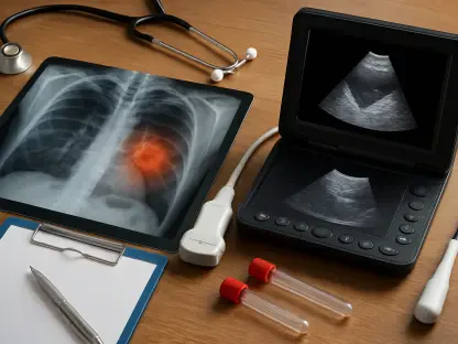

Hospital radiology leaders faced a stark calculation today: how to deliver sharper answers faster without sinking capital budgets into technology that slows throughput or requires specialized workflows, all while keeping radiation dose under control and staff time predictable across stacked schedules and multiple service lines. FDA 510(k) clearance for Philips’ Verida, an AI-enabled detector-based spectral CT, sharpened that equation by promising speed and clarity at a price point meant to pressure photon-counting rivals. The system analyzes X-ray attenuation at different energies to separate materials and enhance contrast, then uses AI to reconstruct up to 145 images per second so exams could be reviewed in roughly 30 seconds. Philips framed Verida as a high-throughput machine that doubles reconstruction speed versus its prior spectral CT, claims dose and image noise reductions around 80%, and slots into standard workstations to avoid a disruptive IT build.
The Race for Premium CT: Speed, Price, and Proof
Spectral CT has long promised better material separation than conventional CT, and Verida leaned into that edge with a detector-first design and AI acceleration tuned for daily volume. By capturing energy-dependent data and feeding it through reconstruction models, the system generated monoenergetic views and material maps aimed at cleaner visualization of iodine, calcium, and soft tissue interfaces. Philips asserted that AI denoising and reconstruction routines cut dose and noise by roughly 80% while doubling reconstruction speed compared with its previous spectral platform, which in turn supported an estimated capacity of up to 270 exams across a 16-hour day. If borne out in practice, those throughputs mattered for emergency departments, oncology pipelines, and cardiac programs where minutes dictated bed flow and contrast timing. The headline, though, still came with an asterisk: these were company claims awaiting broad, independent validation.
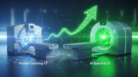
The competitive backdrop grew more pointed. Siemens Healthineers secured clearance for photon counting in 2021, and GE HealthCare followed with a system cleared in March 2026, elevating detector innovation as the new performance benchmark. Photon counting promised superior energy resolution, potential gains in spatial resolution, and cleaner multi-material separation, benefits attractive for small-vessel assessment, lung imaging, and micro-calcification detail. Yet such systems typically carried roughly twice the list price Philips cited for Verida, adding pressure on budgets, service contracts, and siting decisions. Philips countered that routine, high-volume environments favored reconstruction speed, cost efficiency, and workflow continuity, and said it planned to enter spectral photon counting when the technology aligned with high-throughput clinical use. Verida’s pitch hinged on delivering spectral results quickly on standard workstations, minimizing IT complexity while keeping radiologists inside familiar reading patterns.
What Matters Next: Decisions for Buyers
Procurement teams weighed options through a pragmatic lens that balanced capital cost, workflow gains, and measurable clinical impact. Verida’s list price ranged from €1 million to €2 million, with a midpoint near €1.5 million, positioning it as a premium CT that undercut photon-counting alternatives that often landed near double that spend. Total cost of ownership then rested on tube life, service coverage, training, and AI software updates, plus knock-on savings from fewer rescans if noise suppression and dose reductions held up. Regulatory milestones added confidence: a CE mark preceded FDA clearance, and an initial installation in Madrid was underway as of February, providing an early site for workflow insights. Still, the decisive factor remained proof. Health systems that negotiated enterprise imaging contracts, standardized protocols, and reading room ergonomics typically insisted on side-by-side studies, dose registry tracking, and time-to-diagnosis metrics before codifying new scanners into care pathways.
Translating claims into policy required rigor, and the way forward had been clear. Buyers should have commissioned pilot deployments that measured door-to-report time in chest pain workups, staging accuracy in oncology, and repeat-scan rates in bariatric cohorts where dose and noise most stressed image quality. Institutions should have specified structured reporting fields for spectral outputs, integrated AI versioning into PACS governance, and required vendor-supported training that synchronized technologists and radiologists on reconstruction presets. Contract clauses should have tied payments to uptime, reconstruction throughput, and software update cadence, while academic partners should have registered prospective, head-to-head studies comparing photon counting and detector-based spectral CT on lesion conspicuity, iodine quantification, and dose. By anchoring decisions to these steps, health systems had reduced risk, captured operational wins, and positioned their fleets to evolve from 2026 to 2028 without locking into a single detector doctrine.