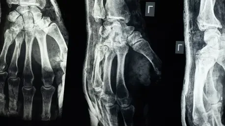

The fragility of AI performance is highlighted by the dramatic drop in sensitivity when a model is applied to data from a source not included in its training set. Primary bone tumors represent one of the most daunting diagnostic hurdles in modern musculoskeletal radiology due to their extreme rarity and the subtle ways they manifest on conventional X-rays. Missing these lesions can lead to catastrophic consequences for patients, yet the nuance required for detection often eludes even the most seasoned radiologists during routine screenings. To bridge this critical gap, a recent study has introduced a biomimetically inspired artificial intelligence framework that prioritizes clinical transparency over the inflated statistics commonly seen in tech marketing. By focusing on honest performance metrics, this research aims to provide a reliable second set of eyes that understands the stakes involved in oncological diagnostics. This shift in focus toward real-world utility ensures that the technology serves as a helpful assistant rather than a distraction in the high-pressure clinical workflow.
The Human-Inspired Workflow: A Cognitive Approach to Architecture
The development of high-performance medical AI requires a shift away from traditional computer vision methods toward architectures that reflect the nuance of clinical practice. Rather than treating bone tumor detection as a standard object identification task, the latest research adopts a biomimetic strategy that prioritizes the way an expert actually interprets a radiograph. This philosophy acknowledges that medical images are not just arrays of pixels but complex biological maps where subtle deviations can indicate life-altering pathologies. By structuring neural networks to follow a logical, stepwise diagnostic path, engineers can create tools that are more intuitive and less prone to the errors associated with “black-box” systems. This evolution in design is particularly crucial for rare conditions like primary bone tumors, where the scarcity of examples makes it difficult for standard models to learn effectively. A human-centric approach to architecture not only improves accuracy but also aligns the technology with the existing expertise of radiologists.
Cognitive Modeling: The Image-Level Classifier
The AI system was meticulously designed to mirror the cognitive workflow of a human radiologist, who typically scans an image for a general impression before hunting for specific abnormalities. The first stage of this biomimetic pipeline consists of an image-level classifier that functions as a sophisticated gatekeeper, determining if a radiograph appears suspicious or normal. This component utilizes an EfficientNetV2-S backbone, which was initially pretrained on the MURA dataset—a massive repository of musculoskeletal radiographs—and then fine-tuned using the Bone Tumor X-ray Repository. The fine-tuning process employed a specialized two-phase unfreezing schedule, allowing the neural network to release its layers gradually. This methodical approach ensures that the model preserves broadly learned medical features while adapting its focus to the highly specific and often faint signatures left by primary bone tumors. By establishing this initial global impression, the system creates a robust foundation for more detailed analysis.
Targeted Localization: The Lesion Detection Phase
Following the initial assessment, the second stage of the pipeline introduces a lesion detector that only activates if the classifier flags a radiograph as abnormal. By implementing this hierarchical gating mechanism, the system effectively avoids running complex localization algorithms on healthy bone structures, which significantly reduces the frequency of false marks that can clutter a diagnostic report. This architecture ensures that the AI remains computationally efficient while focusing its detection capabilities only on the most relevant cases, effectively mimicking the directed attention of a human expert. The use of a YOLOv8 object detection model for the second stage allows for precise localization of suspicious areas, marking them clearly for the radiologist’s review. This balanced approach not only improves the speed of the diagnostic process but also minimizes the cognitive load on the physician, ensuring that the technology acts as a true aid in identifying life-threatening conditions without overwhelming the user.
Computational Efficiency: The Gating Mechanism
One of the primary advantages of this two-stage structure is its ability to suppress false positives by filtering out images that do not exhibit any signs of pathology. In a typical clinical setting, a high volume of radiographs may be completely normal, and an AI that flags even a small percentage of these correctly can lead to “alarm fatigue” among medical staff. The gating mechanism addresses this by ensuring that the localization model never even sees images that the initial classifier deems safe. This logic prevents the detector from finding phantom tumors in the natural textures of healthy bone or in the artifacts common to X-ray imaging. By reducing the search space to only those images with a high probability of disease, the system achieves a much cleaner output. This refinement is essential for the practical adoption of AI, as it respects the limited time of the radiologist and ensures that every notification generated by the software warrants serious attention and review.
Specialized Training: Fine-Tuning and Unfreezing
The technical success of the model relied heavily on a sophisticated training strategy that moved beyond simple supervised learning. By utilizing a two-phase unfreezing schedule, the researchers were able to carefully control how the neural network adapted to the specific characteristics of bone tumors. In the first phase, only the final layers of the model were trained, allowing the system to learn the classification task without disrupting the foundational medical knowledge it gained during pretraining. In the second phase, earlier layers were gradually unfrozen, enabling the network to refine its understanding of the subtle textures and edges associated with bone lesions. This granular approach to training is particularly effective when working with relatively small medical datasets, where over-fitting is a constant risk. By preserving the general features of skeletal anatomy while sharpening the model’s focus on tumor-specific patterns, the researchers created a system that is both robust and highly specialized.
Methodological Rigor: Performance Realities and Ethical Standards
Rigorous scientific inquiry in the realm of medical imaging necessitates a departure from the superficial metrics that often dominate the broader technology industry. For an AI tool to be truly effective in a hospital, it must be evaluated using standards that reflect the actual pressures and requirements of a clinical environment. This means moving beyond simple accuracy percentages and toward a more comprehensive understanding of how a model behaves when faced with real-world constraints. The use of advanced evaluation frameworks allows researchers to simulate the diagnostic process more accurately, identifying potential weaknesses before a tool is ever used on a patient. By establishing a culture of methodological rigor, the research community can ensure that new developments are not just statistically significant but clinically transformative. This dedication to honesty and transparency is what separates a experimental prototype from a reliable medical device.
Clinical Honesty: Evaluation Standards and Integrity
To ensure the results were grounded in reality, the researchers utilized the Bone Tumor X-ray Repository and maintained a strict level of methodological discipline throughout the testing phase. A critical aspect of this study was the decision to lock all decision thresholds on the validation data before ever exposing the model to the held-out test set. This practice stands in sharp contrast to many contemporary AI studies that retroactively adjust their sensitivity settings to achieve the most impressive numerical outcomes, which often leads to inflated performance that cannot be replicated in a live clinical setting. By adhering to these rigid standards, the team provided a more transparent and reliable baseline for understanding how computer-aided detection tools would actually perform inside a hospital. This commitment to data integrity ensures that the technology is judged by its practical utility rather than its potential for marketing, fostering a higher degree of trust among professionals who depend on accurate diagnostic tools.
Statistical Accuracy: Analyzing Performance Metrics
Performance metrics gathered during the study revealed that the AI is particularly adept at identifying malignant tumors, which often cause more aggressive and visible structural changes in the bone than benign lesions. At a primary operating point of only 0.02 false marks per image, the system achieved a lesion-level sensitivity of nearly 65 percent, a figure that climbed even higher when the false-alarm budget was slightly expanded. While these numbers might appear lower than the sensationalized accuracy rates found in general technology news, they represent a high level of precision under the strict constraints required for patient safety and clinical reliability. The discrepancy in performance between malignant and benign cases highlights the inherent difficulty of the task, yet the system’s ability to flag the most dangerous tumors remains a significant achievement. These results provide a realistic perspective on the current state of neural networks in radiology, emphasizing the importance of balancing sensitivity with practical limitations.
Sensitivity Risks: The Fragility of Generalization
One of the most revealing aspects of this research was the implementation of a leave-one-center-out experiment, which tested how the model performed when data from a specific hospital was entirely excluded from its training set. When the largest data source was removed, leaving the model to rely on a significantly smaller pool of images, the system’s sensitivity dropped drastically from over 66 percent to less than 29 percent. This dramatic decline underscores a major challenge in the field of medical artificial intelligence: a system that has been perfected in one facility may fail to maintain its accuracy when deployed in another. Such variations are often caused by differences in imaging equipment, patient demographics, and even the specific radiographic techniques used by local technicians. This finding serves as a sobering reminder that the success of these models is deeply tied to the diversity of the data they consume, and a lack of varied training material can lead to a fragile system that is unable to generalize.
Building Confidence: Transparency and Future Steps
In the final analysis, the research established that while AI could not yet replace human intuition, it functioned as a vital safeguard when integrated into a mature evaluation culture. The move toward Free-response Receiver Operating Characteristic analysis provided a more honest blueprint for how these tools were judged, moving away from abstract metrics and toward practical clinical utility. Future implementations should focus on creating multi-center validation protocols to address the data fragility identified during the testing phase. Hospitals were encouraged to begin establishing standardized data-sharing agreements that would allow for more diverse training sets, which would ultimately improve the sensitivity of these systems across different imaging platforms. By prioritizing clinical transparency and rigorous testing, the field moved a step closer to ensuring that rare bone tumors were caught in their earliest stages. The study concluded that the path forward required a commitment to honesty in performance reporting.