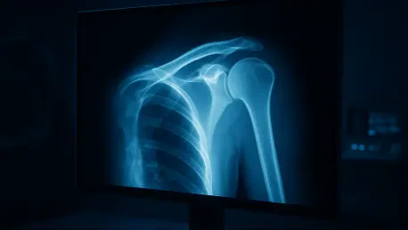

While Magnetic Resonance Imaging serves as the definitive tool for diagnosing tendon damage, its high cost and limited availability frequently delay treatment for patients in resource-constrained environments. Consequently, the clinical landscape of musculoskeletal medicine is defined by a tension between diagnostic necessity and economic reality. Rotator cuff tears represent a primary cause of chronic shoulder pain and physical disability, affecting millions of individuals globally. While MRI is universally recognized as the gold standard for soft-tissue injuries due to its superior contrast and resolution, its implementation is hampered by long wait times and high prices. This leaves the plain shoulder X-ray as the first-line diagnostic tool. However, the inherent limitation of X-rays—their inability to clearly visualize soft-tissue structures like tendons—means that identifying rotator cuff damage from a radiograph is an exercise in subtlety that often eludes even seasoned radiologists. To address this diagnostic gap, researchers from Xinjiang Medical University developed a sophisticated artificial intelligence framework titled WEM-Mamba, or Wavelet-Enhanced MedMamba. This new model seeks to extract invisible diagnostic signals from standard shoulder X-rays by merging advanced state space modeling with classical frequency domain analysis.
Addressing the Spatial Bias in Medical Imaging
The fundamental problem with applying standard deep learning to medical imaging is the “spatial-only” bias found in conventional systems. Most standard architectures, including Convolutional Neural Networks and Vision Transformers, treat images primarily as grids of intensity values in the spatial domain. While these models are excellent at recognizing shapes and local patterns, they often overlook the rich frequency-based information embedded in the texture of an image. In a shoulder X-ray, the clues to a rotator cuff tear are not obvious structural breaks but rather subtle changes in the textural signature of the soft tissue shadows and bone interfaces. By ignoring these nuances, traditional AI may miss the early signs of degeneration. The Urumqi-based research team posits that frequency domain analysis is the key to unlocking these hidden signatures. Digital images can be mathematically decomposed into different frequency bands where high-frequency components represent sharp edges and fine details, while low-frequency components represent broader anatomical structures and transitions.
By analyzing these bands separately and then integrating the findings, the AI can theoretically detect textural irregularities that would be smoothed over in a traditional spatial analysis. This multi-scale perspective mirrors how human experts might interpret complex visual data if they could perceive microscopic variations in pixel density. Traditional algorithms often prioritize macro-structures, but for conditions like rotator cuff tears, the macro-structure shown on an X-ray might appear normal to the naked eye. The innovative approach utilized in this research involves training the model to prioritize micro-textural shifts that indicate underlying pathology. By focusing on the interplay between different frequency bands, the framework identifies patterns associated with indirect signs of tendon damage, such as cortical irregularities or cystic changes in the humerus. This method transforms a simple radiograph into a data-rich source that goes beyond mere bone visualization. The shift from pure spatial processing to a dual-domain approach represents a significant evolution in how machine learning interprets standard diagnostic imagery.
The Innovative Architecture: Mamba Meets Wavelets
Built upon State Space Models, the MedMamba architecture, and the Haar Wavelet Transform, the WEM-Mamba framework addresses the efficiency limitations of current medical AI. For several years, Transformers have dominated the landscape due to their attention mechanisms, which allow models to understand complex relationships within an image. However, Transformers suffer from quadratic scaling, meaning computational requirements grow exponentially with image resolution. This makes them difficult to deploy on standard clinical hardware found in most community hospitals. The Mamba architecture utilizes State Space Models to process data sequences with linear scaling. This provides the global context of a Transformer but with the speed and efficiency necessary for real-world medical environments. By maintaining a linear computational footprint, the model ensures that high-resolution diagnostic tasks can be performed quickly without requiring specialized, high-cost server clusters. This architectural choice makes the system an ideal candidate for immediate bedside application in diverse settings.
The most significant technical contribution of this research is the Wavelet-Enhanced SS-Conv-SSM module, which integrates advanced mathematics with deep learning. Rather than simply feeding a flat image into the network, this module employs the Haar wavelet transform to split the image into four distinct sub-bands. These bands represent low-frequency approximations along with horizontal, vertical, and diagonal high-frequency details. This specific decomposition allows the network to inspect the fine-grained textural data of the shoulder joint in isolation from the coarse anatomical data. By isolating these frequency components, the model can apply specialized processing to each, ensuring that subtle cues indicative of soft-tissue damage are not drowned out by more prominent bone structures. This multi-resolution analysis mimics the way a radiologist might adjust contrast or focus to find a subtle diagnosis. The ability to handle these data streams simultaneously allows the model to capture both local textural nuances and the global orientation of the joint structure, providing a comprehensive diagnostic overview.
Hybrid Processing: From Decomposition to Reconstruction
Once the image has been decomposed, the WEM-Mamba framework utilizes a dual-pathway system to ensure maximum information retention during analysis. Convolutional layers are employed to handle local feature extraction, focusing on the immediate relationships between neighboring pixels. Simultaneously, the state space model manages long-range dependencies across the entire radiograph, ensuring that a feature identified in one part of the shoulder is contextualized by the surrounding anatomy. This hybrid approach allows the system to remain sensitive to minute details while maintaining a cohesive understanding of the patient’s skeletal structure. Finally, an inverse wavelet transform is used to reconstruct these enhanced features into a unified representation. This reconstruction step is critical because it ensures that the final classification is based on a holistic understanding of the data. The model does not just look at isolated patches but integrates frequency and spatial information to arrive at a final diagnostic decision that considers all available radiographic evidence.
This complex processing flow addresses the “black box” problem often associated with medical AI by aligning its internal logic with established mathematical principles. The use of inverse transforms allows the model to map its findings back into a spatial context that aligns with the original X-ray image. By doing so, the system ensures that the features it identifies as diagnostic are actually rooted in the physical anatomy of the shoulder. This method prevents the model from relying on digital artifacts or noise that might be present in imaging but carry no actual clinical significance. Moreover, the integration of these features provides a robust baseline for detecting the subtle footprints of rotator cuff tears, which often present as minor shadows or altered bone density. The end result is a highly focused diagnostic signal that stands out from the cluttered background of a standard radiograph. This technical rigor ensures that the system is not merely guessing based on statistical correlation but is instead performing a structured analysis of the radiograph’s content.
Empirical Performance: Results and Clinical Accuracy
The effectiveness of WEM-Mamba was rigorously tested against fifteen other state-of-the-art AI architectures, including established CNNs like ResNet and modern Transformers. The results demonstrated a clear superiority for the wavelet-enhanced approach, with the model achieving an accuracy of 0.8950 and an F1-score of 0.9309. Particularly significant in a medical screening context was the model’s recall rate of 0.9552. In clinical terms, a high recall means the model is exceptionally good at finding true positives, rarely missing a tear when one is actually present. In a triage scenario, missing a diagnosis is often more dangerous than a false alarm, as it delays necessary treatment for a progressive injury. By ensuring that nearly all potential tears are identified for further review, the WEM-Mamba system serves as a reliable safety net for clinicians who might be working under pressure. This performance suggests that AI can effectively augment human expertise in interpreting the subtle textures associated with musculoskeletal damage.
Beyond its raw accuracy, the model’s efficiency was a standout metric during the testing phase. With only 14.92 million parameters and a computational requirement of 2.04 gigafloating-point operations per inference, WEM-Mamba is significantly leaner than many of its competitors. This low computational footprint suggests that the software could be integrated into existing hospital radiology workstations without the need for expensive, high-end GPU clusters. This efficiency is crucial for deployment in community health centers and rural clinics that may not have the budget for massive computing infrastructure. The ability to run high-level diagnostic AI on standard hardware makes it a viable candidate for large-scale clinical rollout. Furthermore, the speed of inference allows for real-time analysis, meaning a physician could receive a diagnostic flag shortly after the X-ray is taken. This rapid feedback loop significantly reduces the time between initial presentation and the start of a concrete treatment plan for the patient.
Strategic Implementation: The Path Forward
The successful implementation of the WEM-Mamba framework pointed toward a new era of proactive diagnostic strategies in orthopedic medicine. Healthcare administrators and technology providers recognized the necessity of integrating such low-footprint AI systems into existing digital imaging workflows to maximize clinical utility. The next strategic phase involved the establishment of standardized protocols for multi-center data sharing to facilitate the model’s global refinement across diverse demographics. Developers focused on expanding the model’s capabilities to include automated severity scoring, which allowed for immediate clinical triaging without waiting for manual radiologist review. This transition facilitated a more streamlined approach to patient care, where the initial radiograph acted as a high-fidelity gatekeeper for more expensive interventions. By adopting these mathematically enhanced models, medical institutions lowered diagnostic costs while significantly improving the speed of treatment delivery for those suffering from chronic pain.
The validation of this technology encouraged a broader movement toward precision-engineered AI that addressed specific clinical bottlenecks through targeted frequency analysis. Researchers moved beyond binary detection to classify specific tear patterns, providing orthopedic surgeons with actionable data for preoperative planning directly from initial screenings. Multi-center trials were launched to ensure the model remained robust against variations in X-ray hardware and technique across different global regions. These initiatives aimed to eliminate the site-specific bias that historically hampered the deployment of diagnostic software. By focusing on the underlying physics of the image rather than just pixel intensity, the framework established a more reliable standard for non-invasive screening. The project eventually demonstrated that sophisticated mathematical modeling could bridge the gap between high-end imaging and accessible primary care. These advancements collectively ensured that the most effective diagnostic insights were available to patients regardless of their local healthcare resources.