
The release of expert-annotated datasets under open-access licenses encourages global collaboration and allows the scientific community to scrutinize and improve diagnostic algorithms. This initiative addresses the staggering burden of knee osteoarthritis, a condition that currently impacts hundreds of millions of individuals worldwide, leading to chronic pain and significant mobility impairment. The disease is characterized by the progressive deterioration of articular cartilage, the specialized tissue that facilitates low-friction movement within the joint. Historically, clinical diagnosis has been a manual, qualitative endeavor, where radiologists assess magnetic resonance imaging scans to identify visible signs of wear. However, the earliest manifestations of osteoarthritis occur at a microscopic scale, often hidden within cartilage layers that are only a few millimeters thick. Detecting these subtle changes requires a level of precision that transcends human visual capacity, making the identification of early-stage degradation a formidable clinical hurdle. The labor-intensive nature of manual segmentation, which involves tracing the boundaries of the cartilage pixel by pixel, further complicates the diagnostic pipeline. This manual approach is inherently subjective and prone to inter-observer variability, which can lead to inconsistent treatment pathways for patients. By automating this process, medical professionals can achieve a more objective and standardized assessment of joint health, ensuring that interventions are based on precise anatomical measurements rather than qualitative estimates.
Overcoming Technical Limitations: Addressing Global Demographic Needs
The development of advanced artificial intelligence for musculoskeletal imaging has long been hampered by the limitations of traditional convolutional neural networks. While these models are effective at identifying localized patterns, they often struggle with long-range dependencies, which refers to the ability to understand how distant anatomical structures relate to one another within a single scan. For thin and complex structures like knee cartilage, an isolated view of pixels is insufficient; the algorithm must understand the broader context of the surrounding bone and joint fluid to distinguish tissue boundaries accurately. Swin-Unet addresses this by utilizing a hierarchical architecture that maintains a global perspective, allowing the system to accurately delineate where cartilage ends and adjacent structures begin. This shift from local convolution to global attention mechanisms represents a fundamental evolution in how computer vision models interpret medical data. By capturing these broader spatial relationships, the model significantly reduces the “noise” and misclassifications that previously plagued automated segmentation efforts, providing a more reliable foundation for clinical decision-making and longitudinal patient monitoring across various stages of joint degeneration.
Beyond these technical hurdles, researchers have identified a critical lack of demographic diversity in existing medical datasets, which often skew heavily toward Western populations. Anatomical nuances and disease manifestation patterns can vary significantly across different ethnicities, meaning that an AI model trained exclusively on one demographic may not perform with the same accuracy when applied to another. To solve this, the introduction of the OAMRI dataset provides a specialized resource reflecting the anatomical characteristics of Asian populations. This focus on inclusive data ensures that diagnostic tools are not only technically sophisticated but also demographically robust. By broadening the scope of available training data, the scientific community can develop algorithms that are globally applicable, reducing health disparities and ensuring that patients in diverse regions receive the same high standard of automated care. This move toward representative data collection is essential for the ethical deployment of artificial intelligence in healthcare, where the goal is to provide equitable outcomes for all individuals regardless of geographic or ethnic background, thereby fostering a more inclusive environment for medical innovation.
Algorithmic Evolution: The Synergy of Transformers and Specialized Data
The technical foundation of this diagnostic breakthrough rests on the innovative Swin-Unet architecture, which marks a departure from traditional pixel-by-pixel processing toward a Transformer-based approach. Originally developed for natural language processing, Transformers utilize self-attention mechanisms to weigh the importance of different parts of the input data relative to one another. In the context of magnetic resonance imaging, Swin-Unet divides a scan into small patches and calculates the relationships between every patch. This allows the model to zoom in on the incredibly thin edges of the femoral and tibial cartilage while simultaneously keeping an eye on the overall load-bearing architecture of the knee. The “shifted window” approach within the Swin architecture is particularly effective, as it allows for cross-window connections that improve the model’s ability to handle the curvilinear shapes of joint tissues. This dual focus on fine detail and broad context is precisely what is required to navigate the complex environment of an osteoarthritic joint, where inflammation and bone remodeling can often obscure the margins of healthy cartilage.
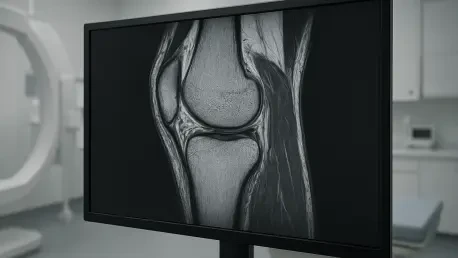
Supporting this sophisticated architecture is the OAMRI dataset, which contains nearly 900 high-resolution, T2-weighted sagittal knee slices. T2-weighted imaging is a specific technique that is highly sensitive to fluid changes and the internal structural integrity of cartilage, making it the ideal medium for detecting early-stage osteoarthritis before physical erosion becomes catastrophic. The dataset focuses on the osteochondral unit, which encompasses the functional interaction between bone and cartilage. By training the AI to recognize the relationship between the femur, tibia, and their respective cartilage layers, researchers have provided the system with a comprehensive understanding of how the knee joint functions as a singular, integrated unit. This holistic approach to data annotation ensures that the AI is not just identifying tissues in isolation but is instead understanding the biomechanical context of the joint. The integration of high-quality, expert-verified data with a cutting-edge Transformer model creates a powerful diagnostic tool that surpasses the capabilities of previous iterations, offering a level of insight that was previously unattainable in standard clinical workflows.
Clinical Validation: Precision Benchmarking and Future Diagnostic Applications
To ensure that the AI model is ready for the rigors of a hospital environment, the research team implemented a series of stringent validation protocols that tested the system’s performance on entirely unfamiliar data. When evaluated using patient-level splits—a testing method where the AI must analyze individuals it has never encountered in its training phase—the model achieved a Dice Similarity Coefficient of approximately 91%. In the field of medical imaging, reaching a score above 90% is a definitive benchmark of excellence, as it indicates that the AI’s digital segmentation nearly perfectly aligns with the manual tracings of human orthopedic experts. This high level of precision suggests that the model is robust enough to handle the natural variations in patient anatomy and the minor differences in imaging quality found in real-world clinical settings. The success of this validation phase demonstrates that the technology has moved beyond theoretical experimentation and is now a viable candidate for integration into the daily routines of radiologists and orthopedic surgeons.
The practical implications of such high-precision automation are transformative, enabling a transition from qualitative observations toward what is known as quantitative radiology. Instead of relying on a doctor’s visual estimate of joint wear, this AI provides hard data on cartilage volume, thickness, and distribution. Such objectivity is invaluable for early detection, as the AI can flag subtle thinning of the cartilage years before a patient begins to experience debilitating symptoms. Furthermore, for surgeons preparing for cartilage repair or partial joint replacement, having a pixel-accurate map of the remaining healthy tissue allows for a highly personalized surgical plan that maximizes the longevity of the repair. In the realm of pharmaceutical development, this technology serves as a tireless auditor for clinical trials, providing the repeatable and precise measurements necessary to determine if a new drug is successfully slowing the progression of the disease. By providing a consistent and objective “second pair of eyes,” Swin-Unet is fundamentally altering the landscape of joint health management and improving the prospects for millions of patients.
Strategic Integration: Advancing Orthopedic Care through Automated Intelligence
The recent advancements in automated segmentation represented a major shift in the way degenerative joint diseases were managed within the healthcare system. By successfully integrating the Swin-Unet architecture with specialized datasets, researchers established a new standard for diagnostic accuracy that previously seemed out of reach. This progress was not merely a technical achievement but a clinical one, as it provided the necessary tools to move away from the subjective grading systems that had dominated the field for decades. The implementation of these AI models facilitated a much faster diagnostic process, allowing medical facilities to handle higher patient volumes without compromising the quality of the analysis. It also proved that deep learning could be adapted to the specific needs of diverse populations, addressing the historical biases that had often marginalized certain groups in medical research. These milestones highlighted the potential for AI to act as a permanent fixture in the orthopedic toolkit, providing a level of consistency that was previously impossible to maintain across different clinics and practitioners.
Moving forward, the focus should shift toward the seamless integration of these diagnostic models into the standard electronic health record systems used by hospitals globally. This would allow for the automatic generation of quantitative reports every time a knee MRI is performed, giving clinicians immediate access to longitudinal data on a patient’s joint health. It is also recommended that further research explores the application of these Transformer-based models to other joints, such as the hip and ankle, where similar degenerative conditions are prevalent. Establishing international standards for the labeling and sharing of medical data will be crucial to maintaining the momentum of these developments and ensuring that AI tools remain transparent and verifiable. By prioritizing the expansion of diverse datasets and refining the algorithmic transparency of these systems, the medical community can ensure that the transition to AI-assisted diagnosis is both ethically sound and clinically effective. These steps will be essential in moving toward a future where joint preservation and early intervention are the standard of care for all aging populations.