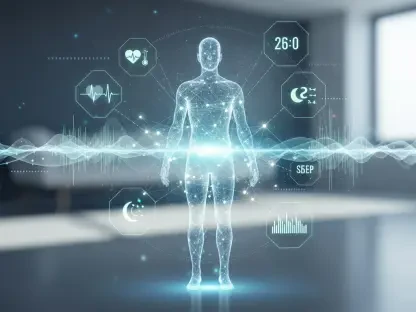

In a rapidly evolving landscape of medical technology, the excitement around artificial intelligence (AI) in breast imaging is palpable, particularly with insights shared by Teri Thomas, CEO of Volpara Health. The potential of AI to revolutionize diagnostic practices, especially in the field of radiology, stands as a paramount discussion point. Thomas strongly believes that future generations will marvel at the fact that radiologists once read images without the aid of AI. This viewpoint sets the stage for a deeper exploration into how AI is poised to transform breast cancer diagnostics, primarily through enhanced accuracy and efficiency.
Volpara Health’s Strategic Acquisition
The Synergy Between Volpara and Lunit
Volpara Health’s recent acquisition by Lunit, a South Korean AI company that specializes in cancer diagnostics, marks a significant step in its commitment to AI’s transformative potential. This strategic move has created a symbiotic relationship, markedly enhancing Volpara’s AI capabilities by merging them with Lunit’s advanced AI tools. The integration of Volpara’s extensive insights into the U.S. healthcare system and Lunit’s cutting-edge technology creates a powerful synergy. This collaboration is expected to substantially elevate the quality of breast cancer diagnostics, making the process more accurate and streamlined.
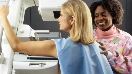
During a visit to Saint Göran Hospital in Stockholm, Teri Thomas witnessed firsthand the impact of integrating AI into mammography. In Europe, the standard procedure for mammogram reading involves two radiologists to ensure unbiased and accurate results. However, due to a significant shortage of radiologists, Saint Göran employs Lunit’s AI as a secondary reviewer. Remarkably, this AI intervention often yields superior results compared to dual human radiologists. Acting as a “second read,” the AI systematically analyzes images, detecting anomalies that might be overlooked in human reviews. This integration underscores the critical role AI plays in mitigating the radiologist shortage while enhancing diagnostic accuracy and efficiency.
Enhancing Diagnostic Accuracy
The collaboration between Volpara Health and Lunit leverages advanced AI tools to boost diagnostic accuracy, particularly in the realm of breast imaging. By employing AI as a secondary read tool, the partnership aims to ensure no anomalies are missed during initial reviews. Radiologists can now benefit from AI’s systematic and unbiased analysis, which renders a dependable second opinion. This methodology notably reduces the chances of oversight, thereby enhancing the overall reliability of breast cancer screenings.
Moreover, employing AI in diagnostic practices addresses the immediate challenge posed by the radiologist shortage. The system ensures consistent and precise secondary reviews, thereby alleviating the pressure on overwhelmed radiology departments. The resulting improvement in diagnostic accuracy is a win-win situation for both patients and healthcare providers, promising more effective and reliable breast cancer detection.
AI Integration in Mammographic Practices
Optimizing Imaging Conditions
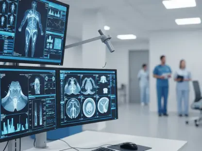
The newfound integration has driven Volpara to incorporate AI into its software solutions, particularly to optimize various aspects of mammographic practices. These include crucial elements such as breast positioning, compression, and radiation dosage, all of which are essential to ensuring high-quality imaging. Thomas emphasized the significance of proper compression; both overcompressing and undercompressing the breast tissue can lead to suboptimal mammograms. AI tools offer precise, data-driven recommendations that assist radiologists in achieving the optimal imaging conditions, leading to high-quality mammograms and reducing the likelihood of errors.
The integration of AI also plays a pivotal role in standardizing imaging practices. By relying on AI-generated guidelines, radiologists can maintain consistent quality in breast imaging across different patients and settings. This consistency is fundamental in ensuring that all patients receive reliable and accurate diagnostics, thereby fostering a more systematic approach to breast cancer screening.
Addressing the Radiologist Shortage
One of the most pressing issues in modern healthcare is the acute shortage of radiologists, which significantly impacts diagnostic practices. The utilization of AI in mammographic practices directly addresses this challenge by providing an efficient and accurate “second read.” By automating the review process through sophisticated AI algorithms, healthcare facilities can manage more cases without compromising the quality of diagnosis. This not only alleviates the burden on existing radiologists but also ensures that patients receive timely and precise screenings, which is crucial for early detection and treatment of breast cancer.
Furthermore, AI’s ability to analyze vast amounts of data quickly results in faster turnaround times for diagnostic results. This efficiency is particularly beneficial in high-volume clinics and hospitals where the demand for radiology services far exceeds the available resources. Consequently, the overall patient experience is significantly enhanced, reducing the anxiety associated with waiting for diagnostic results and fostering a more responsive healthcare system.
The Path Forward
In the rapidly advancing world of medical technology, there’s palpable excitement about the role of artificial intelligence (AI) in breast imaging. This was particularly highlighted by Teri Thomas, CEO of Volpara Health. The potential for AI to revolutionize diagnostic practices, especially within radiology, is a crucial point of discussion. Thomas envisions a future where people will be astonished that radiologists once interpreted images without the assistance of AI. Her perspective opens the door to a deeper exploration of how AI is set to transform breast cancer diagnostics. AI promises to greatly improve the accuracy and efficiency of these processes, leading to earlier and more precise detection of breast cancer. The use of AI can help reduce human error, speed up diagnosis times, and provide personalized care plans for patients. As this technology continues to develop, it holds the promise of not just enhancing existing diagnostic methods but also paving the way for innovations that could fundamentally change how breast cancer is detected and treated.