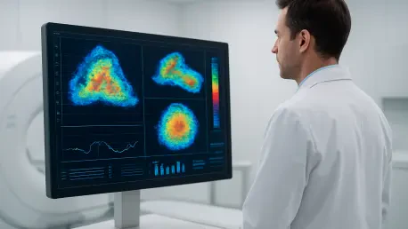

Integrating MRI-derived measurements of myelin density into amyloid PET models allows clinicians to correct signal interference and improve diagnostic accuracy by approximately six percent. This advancement represents a fundamental shift in how modern medicine approaches the diagnosis and treatment of complex physiological conditions. Instead of relying solely on anatomical changes that often appear only after a disease has caused significant damage, physicians are now utilizing molecular imaging to visualize the very biological processes that drive illness. This transition from macro-level observation to micro-level analysis means that healthcare providers can detect the earliest signs of cellular dysfunction, offering a level of precision that was previously unattainable. By leveraging the combined power of high-resolution structural data and sophisticated metabolic tracers, the medical community is effectively moving toward a standard of care where every therapeutic decision is dictated by the unique molecular profile of the individual. This nuanced approach ensures that treatments are not just reactive measures but proactive interventions tailored to the specific cellular environment of each patient, fundamentally altering the trajectory of chronic and acute disease management.
Redefining Early Detection: The Shift Toward Molecular Sensitivity
A significant advancement in the management of neurodegenerative disorders involves the identification of subtle “gray zones” within brain scans that were previously categorized as either normal or abnormal. Rather than adhering to a binary system for amyloid plaque detection, researchers are now focusing on intermediate levels of protein deposition that serve as early warning signs of cognitive decline. These intermediate states represent a critical window for medical intervention, as they often precede the onset of clinical symptoms by several years. By identifying these high-risk profiles during the earliest stages of protein accumulation, clinicians can implement neuroprotective strategies long before irreversible neuronal loss occurs. This focus on the biological spectrum of disease, rather than a simple positive or negative diagnosis, allows for a more granular understanding of how Alzheimer’s and related conditions evolve. It also facilitates more effective screening for clinical trials, ensuring that the participants most likely to benefit from experimental therapies are identified with a degree of accuracy that matches the sophisticated nature of contemporary drug development.
Furthermore, the relationship between amyloid plaques and tau tangles has become a focal point for refining diagnostic timelines and prognostic accuracy. While the presence of amyloid often marks the initial stages of a neurodegenerative process, the accumulation of tau protein is more directly correlated with actual cognitive impairment and the physical destruction of brain tissue. Modern molecular imaging protocols now prioritize the mapping of both proteins to create a comprehensive biological narrative for each patient. By catching these biological shifts at an intermediate stage, healthcare systems are successfully pivoting from the management of advanced dementia to the proactive preservation of brain health. This evolution is particularly crucial for developing longitudinal care plans that adjust as the patient’s biological markers change over time. The ability to distinguish between a stable condition and one that is rapidly progressing allows for a more efficient allocation of resources and a more compassionate, evidence-based approach to patient counseling and long-term care strategy.
Enhancing Diagnostic Accuracy: The Power of Multi-Modal Imaging
To further refine the reliability of neurological assessments, the integration of different imaging modalities has become a standard practice for resolving long-standing technical challenges. One of the most significant hurdles in PET imaging is the interference caused by white matter and myelin density, which can distort the signal and lead to an overestimation of protein deposits. By utilizing high-resolution MRI to measure the specific density of myelin in a patient’s brain, clinicians can now apply mathematical corrections to PET data, ensuring that the resulting images reflect the true biological state of the tissue. This synergy between structural and functional data sources minimizes the risk of false positives and provides a much clearer picture of the disease’s progression. As these multi-modal techniques become more common, they are setting a new benchmark for diagnostic precision, allowing doctors to track minute changes in brain chemistry with a level of confidence that was once hindered by the physical limitations of single-modality hardware.
This collaborative approach to imaging also plays a vital role in long-term longitudinal studies, which are essential for understanding how chronic diseases behave across different populations. When PET and MRI data are harmonized, researchers can track biological changes over several years without the data being obscured by physiological noise or individual anatomical variations. This technical synergy is particularly effective for monitoring the efficacy of new medications, as it allows for the detection of subtle improvements in cellular health that might not be visible on a standard clinical scan. By reducing the risk of data masking, these advanced imaging protocols ensure that the medical community has access to the most reliable information possible to guide future treatment paradigms. This shift toward highly integrated diagnostics is not just an incremental improvement in technology; it is a holistic reorganization of medical data that prioritizes accuracy and longitudinal consistency above all else.
Transforming Oncology: Real-Time Feedback and Theranostic Loops
In the field of oncology, the emergence of theranostics has revolutionized the treatment of aggressive malignancies, such as prostate cancer, by creating a seamless loop between diagnosis and therapy. This approach involves using a specific molecular tracer to identify the location of tumor cells and then using a structurally similar radioactive isotope to deliver a targeted therapeutic dose to those exact sites. Immediately following the administration of the therapy, SPECT/CT scans are utilized to confirm that the medication has successfully bound to the intended targets. This real-time feedback loop serves as a powerful prognostic tool, allowing oncologists to verify the treatment’s distribution and impact almost instantly. If the scan reveals that the therapy is not reaching certain tumor sites, physicians can quickly pivot to alternative strategies, avoiding the delay and toxicity associated with ineffective treatments. This personalized feedback mechanism ensures that the therapeutic regimen is constantly optimized based on the actual behavior of the cancer within the patient’s body.
The use of dual-tracer PET scans has further enhanced this precision by allowing oncologists to map the internal diversity of complex tumors. It is now well-understood that a single tumor can contain multiple populations of cells with varying metabolic characteristics and levels of aggressiveness. By employing different tracers that target distinct biological pathways, such as glucose metabolism and specific protein expression, doctors can create a multi-layered map of the tumor’s landscape. This comprehensive mapping is essential for identifying high-risk “hotspots” that might be resistant to conventional therapies or likely to metastasize. Armed with this detailed biological information, medical teams can design multifaceted treatment plans that address the unique challenges posed by a tumor’s internal heterogeneity. This strategy moves beyond the “one-size-fits-all” model of cancer care, ensuring that even the most complex cases are managed with a level of specificity that prioritizes the eradication of the most dangerous cell populations while sparing healthy tissue.
Pathogen-Specific Diagnostics: A New Era for Infectious Disease
One of the most persistent challenges in managing infectious diseases is the difficulty of distinguishing between a live, active infection and a localized inflammatory response. Traditional imaging techniques often struggle to differentiate these two states because both can appear as areas of increased metabolic activity on a standard scan. However, new molecular tracers are now being utilized to act as “Trojan Horses” that are exclusively absorbed by specific pathogens, such as the bacterium Pseudomonas aeruginosa. These tracers are designed to mimic the essential nutrients or signaling molecules used by the bacteria, allowing them to be pulled directly into the bacterial cells. When these tracers are visualized on a scan, they provide definitive proof of a live infection, enabling clinicians to identify the exact cause of a patient’s symptoms. This high level of specificity is a game-changer for antibiotic stewardship, as it ensures that powerful medications are used only when a confirmed pathogen is present, thereby reducing the risk of resistance.
The transition toward pathogen-specific imaging is a central component of the broader movement from descriptive to mechanistic medicine. Instead of simply identifying where an abnormality exists, molecular imaging answers the critical questions of what the abnormality is and how it is behaving at a cellular level. This ability to see the “mechanics” of an infection or a chronic condition streamlines the entire management process, from initial diagnosis to the final verification of a cure. As these technologies are integrated into standard clinical workflows, they are reducing the period of diagnostic uncertainty that often plagues patients with complex or recurring infections. By providing a clear and immediate identification of the biological culprit, molecular imaging is enabling a more direct and efficient path to recovery. This evolution not only improves individual patient outcomes but also enhances the overall efficiency of the healthcare system by eliminating the need for exploratory treatments and redundant testing.
Future Considerations: Scaling Personalized Molecular Insights
In recent years, the medical community successfully navigated the transition from generalized diagnostic protocols to the sophisticated molecular frameworks that define modern healthcare. Clinicians recognized that the traditional reliance on anatomical observation was insufficient for the complexities of 2026-era pathologies. The widespread adoption of myelin-corrected PET scans and dual-tracer oncology mapping demonstrated a clear commitment to reducing diagnostic error and enhancing patient safety. These advancements were not merely technical milestones; they represented a fundamental change in the philosophy of patient care, where the biological uniqueness of the individual became the primary driver of therapeutic intervention. By prioritizing the quantification of molecular data, health systems managed to significantly reduce the incidence of ineffective treatments and improved the accuracy of long-term prognostic assessments across a wide range of specialties.
Looking forward, the next logical step for the industry involves the standardization of these molecular imaging protocols across diverse clinical settings to ensure equitable access to precision medicine. Health systems must prioritize the specialized training of radiologists and nuclear medicine physicians to handle the increasingly complex data sets generated by multi-modal scans. Furthermore, the integration of artificial intelligence for the automated quantification of molecular markers will be essential for managing the high volume of data produced during longitudinal studies. As these tools become more accessible, the focus should shift toward developing more affordable and stable tracers that can be distributed to regional hospitals and smaller clinics. By expanding the infrastructure for molecular diagnostics, the medical field can ensure that the benefits of personalized medicine are not limited to major research centers but are available to every patient who requires a precise, data-driven approach to their health management.