
The implementation of patient-specific 3D assets helps eliminate the need for on-the-fly adjustments when surgeons encounter unexpected anatomical variations. For decades, the medical community relied on the mental translation of flat, grayscale images into three-dimensional mental constructs, a process that inherently carried risks of spatial misinterpretation. Today, the convergence of advanced computing and additive manufacturing has fundamentally altered this landscape. By converting high-resolution data from diagnostic scans into physical or virtual representations, medical teams now operate with a degree of foresight that was previously unattainable. This transition represents more than just a technological upgrade; it is a paradigm shift in surgical preparation that replaces guesswork with empirical, tactile evidence. As these tools become standard practice, the margin for error narrows significantly, allowing for more conservative incisions, shorter recovery times, and a higher standard of predictable care for complex cases.
The Journey: From Medical Scans to Physical Replicas
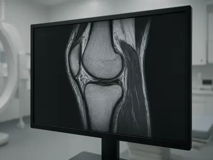
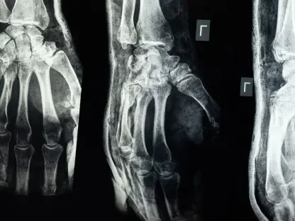
The transformation of raw patient data into a functional surgical tool begins with the meticulous processing of standard diagnostic imagery. While Computed Tomography and Magnetic Resonance Imaging have long been the cornerstones of diagnosis, the resulting images are traditionally viewed as a series of two-dimensional cross-sections. For a surgeon dealing with a rare congenital heart defect or a tumor entwined within a complex vascular network, these slices can fail to convey the true spatial relationships between healthy tissue and pathology. Through the process of segmentation, specialized software isolates specific anatomical regions of interest, allowing engineers to strip away irrelevant data and highlight critical structures. This digital refinement creates a high-fidelity blueprint that serves as the foundation for all subsequent 3D applications, ensuring that every contour and vessel is captured with millimeter-level accuracy before any physical intervention.
Once the digital reconstruction is complete, the data can be leveraged across multiple platforms depending on the specific requirements of the surgical team. In some instances, these models are uploaded into immersive virtual reality environments, enabling surgeons to walk through a patient’s internal anatomy or view it from perspectives that would be impossible during a live operation. Alternatively, these models are sent to high-resolution 3D printers, where they are rendered into physical replicas using materials that mimic the haptic feedback of human bone, muscle, or vascular tissue. These tangible organs can be sterilized and brought directly into the operating room, providing a physical reference point that stays with the surgeon throughout the procedure. This bridge between the digital and physical realms allows for a level of rehearsal that ensures every member of the surgical staff is fully synchronized regarding the patient’s unique geometry.
Precision Planning: Enhancing Intraoperative Efficiency
One of the most significant impacts of 3D modeling lies in the development of bespoke surgical guides and templates that are printed specifically for a single patient’s anatomy. In orthopedic and neurosurgical procedures, where the placement of a screw or the angle of a bone cut is critical, these custom guides are designed to fit perfectly onto the patient’s bone structure. By locking into a unique anatomical position, the guide ensures that surgical instruments, such as drills or saws, follow a predetermined trajectory that has been vetted in a digital simulation. This level of guided precision minimizes the trauma to surrounding healthy tissue and virtually eliminates the risk of misaligned implants. Such technological intervention shifts the focus of the operating room from exploratory decision-making to the efficient execution of a well-defined plan, which is particularly vital during high-stakes maneuvers near the spinal cord or major arteries.
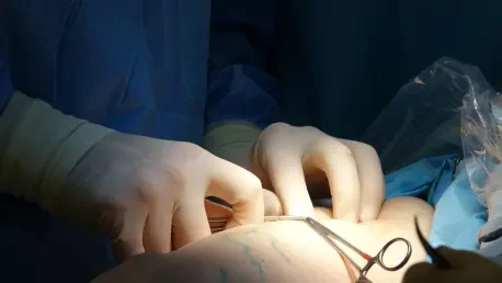
Beyond guiding the actual cuts, 3D printing allows for the pre-operative preparation of permanent hardware, which drastically reduces the time a patient spends under anesthesia. Traditionally, surgeons might have to manually bend titanium plates or rods to match a patient’s bone contour while the patient is already on the operating table, a process that is both time-consuming and labor-intensive. With access to a 3D-printed replica of the patient’s anatomy, these adjustments can be performed in a controlled setting days before the surgery occurs. The hardware is pre-shaped to the exact curvature of the replica, ensuring a perfect fit the moment it is introduced into the surgical site. This advancement not only streamlines the workflow but also preserves the structural integrity of the metal implants by reducing the fatigue caused by repetitive manual bending. Consequently, the entire surgical team can move through the procedure with increased speed and confidence.
Institutional Strategy: Localized Labs and Patient Trust
A pivotal shift in the adoption of this technology is the movement toward establishing centralized 3D printing service lines directly within the hospital infrastructure. Rather than outsourcing the creation of models to distant third-party vendors, which can introduce delays and communication barriers, many institutions are now integrating dedicated laboratories into their own facilities. This proximity allows for a dynamic and iterative design process where clinical engineers and surgeons can collaborate in real-time. When a surgeon identifies a specific anatomical challenge, they can walk down the hall to the lab and work with the engineering team to refine a model or guide until it meets the precise needs of the upcoming procedure. This localized approach ensures that the technology is highly responsive to the urgent timelines often found in acute care settings, where waiting several days for a shipment from an external provider is simply not a viable option for the patient.
Moving forward, the industry successfully transitioned these tools from experimental novelties to essential clinical standards. Hospitals that invested early in 3D infrastructure provided a blueprint for how to balance technological advancement with practical financial sustainability. The widespread adoption of these methods suggested that the future of surgery relied on a holistic approach where data-driven planning and tactile execution were inseparable. Clinicians recommended that medical institutions prioritize the training of specialized staff to manage these assets, ensuring that the technology remained accessible and effective across all surgical disciplines. As the field evolved, the focus shifted toward the automation of segmentation and the use of even more durable, bio-compatible materials for long-term implants. These advancements collectively reinforced the importance of personalized medicine, ultimately proving that the most effective surgical outcomes were achieved when the treatment was as unique as the patient’s own anatomy.