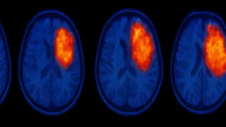

Integrating blocking studies with single-cell analysis provides a methodology to subtract artifacts and reveal the true biological landscape of a brain tumor. Glioblastoma remains one of the most formidable challenges in modern oncology due to its aggressive nature and tendency to infiltrate healthy brain tissue far beyond what is visible to the naked eye. For years, clinicians have relied on Magnetic Resonance Imaging to map these malignancies, yet this standard tool often fails to define the true boundaries of the disease. MRI primarily highlights anatomical changes and fluid accumulation rather than direct cellular activity, which frequently leads to incomplete surgical resections and a high rate of recurrence. To bridge this diagnostic gap, researchers have increasingly turned to Positron Emission Tomography scans, which offer a window into the metabolic and molecular landscape of the malignancy. Despite this promise, a significant scientific debate has emerged regarding the reliability of the signals produced by specific radiotracers. The core of the controversy lies in whether the bright signals seen on a scan represent true tumor biology or are simply an artifact of a compromised blood-brain barrier. In glioblastoma, the protective barrier often breaks down, potentially allowing tracers to leak passively into the tissue and create a false impression of tumor density.
Validating Molecular Targets in Brain Tumor Imaging
The Significance of TSPO and Radiotracer Specificity
The focus of recent molecular investigation is the 18-kDa translocator protein, a marker highly expressed in both glioblastoma cells and the surrounding immune cells that the tumor recruits for its own survival. Radiotracers such as [18F]GE-180 were designed to bind specifically to this protein, serving as a biological beacon for the tumor’s presence even in areas where the brain structure looks relatively normal. However, skeptics have long argued that in late-stage disease, where the blood-brain barrier is heavily damaged, these tracers might just be flooding into the brain through vascular gaps. This passive accumulation could lead to an overestimation of tumor spread, making it difficult for surgeons to know which areas require aggressive intervention and which are merely showing signs of vascular leakage. Determining whether the signal is a result of active protein binding or passive accumulation is essential for accurate diagnosis and the effective planning of personalized therapies.
The debate over radiotracer specificity has slowed the clinical adoption of TSPO-PET imaging, as medical professionals require certainty that the data they see reflects cellular pathology rather than simple plumbing issues within the brain’s blood vessels. If the “glow” on a PET scan is largely driven by a leaky barrier, then the tracer serves as little more than a glorified contrast agent similar to those used in MRI. This specificity problem is particularly acute in glioblastoma because the tumor actively destroys the tight junctions of the blood-brain barrier as it grows. Consequently, the imaging community has been divided, with some advocates pushing for the tracer’s widespread use based on its high tumor-to-background ratio, while others remain cautious, fearing that the lack of clear differentiation between binding and leakage could lead to diagnostic errors. Resolving this uncertainty is paramount to advancing neuro-oncology and ensuring that molecular imaging can fulfill its potential as a precision medicine tool.
Experimental Models and the Blocking Technique
To resolve this long-standing uncertainty, a research team at LMU University Hospital utilized advanced mouse models to distinguish between specific and non-specific PET signals with unprecedented precision. By employing the SB28 glioblastoma line, which mimics the human condition by growing within a functional immune system, the scientists were able to observe the tumor’s interaction with the brain environment in real time. The key to their approach was a sophisticated “blocking” technique, where a massive dose of a non-radioactive ligand was administered to saturate all available protein binding sites before the radioactive tracer was introduced. This method effectively “shuts off” the specific biological signal, leaving behind only the background noise caused by non-specific processes. Any radioactivity detected by the scanner after this saturation must be the result of passive leakage through the compromised barrier, providing a clear quantitative measure of the tracer’s true performance.
The rigor of this experimental design allowed the researchers to isolate the impact of vascular permeability from the actual density of the tumor cells. By comparing the scans of mice that received the blocking dose with those that did not, the team could calculate exactly what percentage of the PET signal was tied to the TSPO protein. This type of controlled study is difficult to perform in human subjects, as it requires the administration of very high doses of non-radioactive compounds, but the mouse model provided the perfect laboratory for such a fundamental investigation. The findings from these experiments have provided the first definitive data points in the debate over radiotracer behavior, moving the conversation away from theoretical speculation and toward empirical evidence. By isolating the background noise, the study has paved the way for a more nuanced understanding of how molecular imaging interacts with the complex physiology of an aggressive brain tumor.
Quantifying Signal Accuracy and Vascular Permeability
Shifting Dynamics of Signal Specificity
The findings from the LMU study revealed that the accuracy of a PET scan is not a fixed value but rather a dynamic property that changes as the disease progresses. In the early stages of tumor development, the research showed that approximately 72% of the PET signal was specific, representing genuine binding to the target TSPO protein. This indicates that when the tumor is still relatively small and the blood-brain barrier is mostly intact, the scan provides a highly reliable map of the malignancy’s molecular environment. However, as the cancer advances and the barrier becomes more porous, the proportion of specific signal drops significantly, reaching roughly 59% in later stages. While the majority of the signal remains biologically relevant even in advanced cases, the “noise” created by the leaky barrier becomes increasingly prominent over time, which could potentially complicate the interpretation of scans in patients with large, high-grade glioblastomas.
This shifting balance between signal and noise suggests that clinicians must be mindful of the timing of the scan and the stage of the disease when interpreting PET data. The increase in non-specific signal in late-stage tumors is a direct reflection of the chaotic vasculature that characterizes aggressive glioblastoma. As the tumor recruits new blood vessels to fuel its growth, those vessels are often malformed and “leaky,” allowing the radiotracer to pool in the extracellular space. Despite this increase in background interference, the absolute signal within the tumor still remained significantly higher than in healthy brain tissue, suggesting that the tracer remains a valuable diagnostic tool throughout the course of the disease. The challenge lies in developing software algorithms or clinical protocols that can account for this rising baseline of leakage, ensuring that the final image presented to the surgeon is as clean and accurate as possible.
Cellular-Level Verification via scRadiotracing
To further validate these findings, the research team utilized an innovative technique known as scRadiotracing to observe the tracer’s behavior at the individual cellular level. This process involved dissociating the tumor tissue into its component cells and analyzing them using flow cytometry to determine exactly where the radioactive molecules were landing. The results were remarkably clear: when the TSPO binding sites were blocked with a cold ligand, the uptake of the tracer inside both tumor cells and immune cells plummeted by 95% and 98%, respectively. This proved beyond a doubt that the tracer is exceptionally effective at finding and binding to its biological target once it successfully reaches the cell. The “specificity problem” identified in earlier studies is therefore not a failure of the tracer’s chemical affinity, but rather an extracellular phenomenon where free-floating molecules pool in the gaps between cells.
By looking past the general “glow” of the scan and focusing on the cellular mechanics, the researchers were able to confirm that the tracer does not bind non-specifically to other proteins or tissues within the brain. This cellular-level verification is a critical piece of the puzzle, as it demonstrates that the biological signal captured by the PET scan is honest and representative of the tumor’s cellular makeup. The high degree of intracellular specificity means that if a way can be found to mathematically subtract the extracellular leakage, the resulting image would be a near-perfect representation of the tumor’s density and inflammatory state. This insight is driving new efforts to refine imaging protocols, as it confirms that the fundamental chemistry of tracers like [18F]GE-180 is sound. The focus has now shifted from questioning the tracer itself to understanding the physical environment of the brain and how it influences the delivery of molecular markers.
Future Directions for Clinical Interpretation
Reconciling Biological Signals and Technical Artifacts
The research conducted at LMU University Hospital successfully bridged two opposing scientific viewpoints by validating concerns about vascular leakage while simultaneously confirming the fundamental value of TSPO-PET imaging. It demonstrated that while a broken blood-brain barrier does introduce a degree of artifactual signal, the majority of the data remains rooted in the actual molecular activity of the tumor and its microenvironment. This reconciliation allows the medical community to move forward with a more balanced perspective, recognizing that PET scans are not infallible but are significantly more informative than traditional anatomical imaging alone. By understanding the physical relationship between barrier permeability and tracer accumulation, clinicians can now begin to develop more sophisticated interpretation models that account for the shifting specificity seen in late-stage glioblastomas.
This study established that the “truth” of a medical image is often a composite of several different physical and biological processes. In the early stages of glioblastoma, a TSPO-PET scan serves as a high-fidelity map of the tumor’s landscape, while in later stages, it becomes a dual indicator of both cellular density and vascular failure. Neither of these pieces of information is useless; in fact, knowing where the blood-brain barrier has failed can be just as important for drug delivery planning as knowing where the tumor cells are located. The key takeaway for the field of neuro-oncology is the necessity of a nuanced approach to imaging, where the biological “signal” and the technical “artifact” are both recognized and managed. This transition from a binary debate over specificity to a quantitative understanding of imaging dynamics represents a major step forward for the industry in 2026.
Navigating Diagnostic Challenges: Actionable Pathways for Neuro-Oncology
Moving forward, the implementation of blocking-based methodologies and single-cell verification techniques set a new standard for the validation of any new radiotracer entering the clinical pipeline. The Munich study showed that it was possible to create a mathematical blueprint for “subtracting” the noise of a leaky barrier, a strategy that should now be applied to other tracers and different neurological conditions. For instance, in diseases like Alzheimer’s or multiple sclerosis, where the blood-brain barrier is also known to be compromised, similar studies could help clarify whether the signals seen in those scans are truly representative of neuroinflammation or are simply products of vascular permeability. Applying this rigorous framework across the board will ensure that the next generation of molecular imaging tools is both highly sensitive and highly specific, regardless of the physical state of the patient’s brain vasculature.
In the clinical setting, the most immediate next step involves the integration of dynamic PET protocols that can measure the rate of tracer accumulation over time. By analyzing the “wash-in” and “wash-out” phases of the tracer, radiologists may be able to distinguish between the fast-moving molecules that bind to cells and the slower-moving molecules that pool in the extracellular space. This approach, combined with the insights gained from the scRadiotracing experiments, offers a practical path toward cleaner, more accurate diagnostic images. Ultimately, the goal was to ensure that the visual information used to guide surgical resections and radiation therapy is as accurate as possible. By accounting for the reality of blood-brain barrier leakage rather than ignoring it, the medical community found a way to enhance the precision of glioblastoma treatment and improve the long-term outlook for patients facing this devastating disease.