
Every second that passes during an acute ischemic stroke translates into the loss of nearly two million neurons, making the speed of medical intervention the single most important factor in determining a patient’s long-term quality of life and recovery prospects. Cercare Medical recently achieved a major regulatory milestone with the FDA 510(k) clearance of its cone-beam computed tomography (CBCT) perfusion software, a move that signals a significant shift in how hospitals manage stroke emergencies. This technology is designed to transform acute stroke management by bringing advanced imaging directly into the neurointerventional suite, where clinicians often struggle with limited visual data. By allowing for high-quality, real-time brain tissue assessment while the patient is on the treatment table, the solution addresses a major gap in current care. It offers the potential for faster and more accurate decision-making during time-sensitive procedures, ensuring that the critical “time is brain” window is maximized for the patient’s benefit.
Modernizing the Stroke Care Pathway
Streamlining Workflows: Faster Intervention at the Point of Care
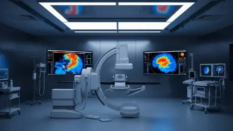
The traditional journey for a stroke patient is often fragmented and fraught with delays, requiring multiple transfers between the radiology department and the angiography suite for different stages of diagnosis and treatment. Cercare’s CBCT perfusion technology removes these logistical hurdles by allowing clinicians to generate CT-quality perfusion maps using standard C-arm systems already present in the operating room. This “point-of-care” capability supports an emerging “direct-to-angio” model where patients can bypass preliminary imaging steps in the emergency room and head straight to the intervention room. Since every minute saved in stroke treatment preserves millions of neurons and improves the chances of a full recovery, reducing these transfers is a critical step in modernizing the survival and recovery rates for patients globally.
Furthermore, the implementation of this software streamlines the communication between different medical teams, as the data is generated and analyzed in the same room where the surgery occurs. By eliminating the need to wait for radiology reports from a different floor, the interventional team can maintain a continuous workflow, reducing the cognitive load on surgeons and nurses. The ability to verify the success of a procedure immediately without moving the patient ensures that the clinical pathway is as efficient as possible. This approach naturally leads to a more integrated environment where the neurointerventional suite functions as a comprehensive diagnostic and therapeutic hub. This shift reduces the overall time from hospital arrival to reperfusion, which is the most reliable predictor of positive neurological outcomes in acute cases.
Advanced Biomarkers: Measuring Real-Time Metabolic Brain Health
Beyond simple blood flow measurements, this software provides a comprehensive look at the brain’s metabolic state through the use of advanced biomarkers that were previously unavailable in the interventional suite. In addition to standard metrics like cerebral blood flow and volume, the platform measures complex factors such as oxygen extraction and capillary efficiency. These physiological indicators offer a more detailed view of the “penumbra,” which is the at-risk brain tissue that surrounds the core of the stroke and can still be saved if blood flow is restored quickly. By understanding the metabolic health of these cells in real time, medical teams can better judge the viability of the tissue and the likely success of the treatment, moving beyond anatomical imaging into functional assessment.
This level of detail allows for a personalized approach to stroke care, as no two patients exhibit the same pattern of tissue decay or vascular collateralization. For instance, a patient with robust collateral circulation might have a larger salvageable penumbra even hours after the initial onset of symptoms, while another might experience rapid tissue death. The software’s ability to map oxygen extraction helps clinicians identify which areas are under the most stress and which are already beyond saving. This data-driven insight prevents unnecessary risks during a procedure and helps manage expectations for recovery. By providing a clear picture of the metabolic landscape, the technology ensures that every decision made on the table is backed by the most current and specific physiological data available.
Improving Diagnostic Precision and Clinical Outcomes
Overcoming Limitations: Capillary-Level Deficits and the No-Reflow Phenomenon
A significant challenge in stroke care is the “no-reflow” phenomenon, where a major vessel is successfully opened, but the brain tissue still does not receive adequate blood because of microvascular damage. Traditional digital subtraction angiography can show that a large vessel is clear of blockages, but it often fails to detect deficits at the microscopic capillary level that prevent oxygen from reaching the neurons. Cercare’s solution fills this diagnostic gap by providing real-time data on tissue-level perfusion while the procedure is still in progress. This allows interventionalists to identify persistent issues or missed blockages that are too small for standard angiography to see, enabling them to adjust their surgical approach immediately before the patient is removed from the table.
The clinical utility of this technology was confirmed through a rigorous multi-reader study at Bern University Hospital in Switzerland, which validated its effectiveness in a real-world setting. During the trial, thirteen neuroradiologists reviewed the data to confirm the accuracy and consistency of the CBCT perfusion maps compared to traditional imaging standards used in radiology departments. The software was built on a vendor-neutral “Vascular Model” engine, which ensured that the maps generated in the intervention room looked familiar to doctors who were used to working with standard MRI or CT scans. This consistency proved vital for ensuring that the technology was easily adopted into existing hospital workflows without requiring extensive retraining for the staff.
Strategic Partnerships: Setting a New Global Standard for Care
In a move to accelerate global adoption and improve patient accessibility, Cercare partnered with Siemens Healthineers to integrate its software directly with the Syngo DynaCT system. This collaboration provided an end-to-end solution for stroke care, from the initial assessment of the patient to the detection of complications during surgery. By bringing this level of physiological detail into the operating room, the partnership aimed to set a new standard for neurointerventional radiology across the healthcare industry. This shift toward personalized, data-driven treatment ensured that successful vessel clearing translated into actual tissue recovery. The integration allowed hospitals to maximize their existing hardware investments while significantly upgrading their diagnostic capabilities.
The implementation of these tools throughout 2026 reflected a broader industry trend toward high-precision, real-time feedback loops in the operating room. Surgeons used the enhanced imaging to confirm that blood flow reached the smallest vessels, reducing the rate of secondary strokes and improving long-term health outcomes. These advancements shifted the focus of neurointervention from simple mechanical revascularization to comprehensive tissue restoration. As more facilities adopted these vendor-neutral platforms, the disparity between radiology-based diagnostics and interventional therapeutics began to close. The clinical community recognized that the future of stroke care rested on the ability to treat and verify results simultaneously, ensuring that every patient received the most informed care possible.