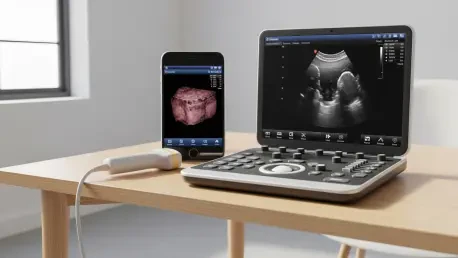

Early detection remains the most critical factor in surviving a breast cancer diagnosis, yet current medical protocols often leave patients vulnerable during the long stretches between annual hospital visits where aggressive tumors can emerge undetected. Researchers at MIT have successfully engineered a portable, operator-independent ultrasound system that challenges the necessity of bulky clinical equipment and highly specialized training for high-quality diagnostic imaging. This breakthrough allows for frequent, accessible screenings, effectively bridging the gap between standard yearly mammograms and the real-time health needs of high-risk individuals. By integrating sophisticated transducer technology into a wearable or handheld format, the team has created a tool that transforms the patient from a passive recipient of care into an active participant in their own health monitoring. This shift toward localized diagnostics represents a fundamental change in how the medical community approaches early intervention and long-term surveillance.
Mitigating Risks of Aggressive Interval Cancers
Traditional screening schedules typically rely on annual mammograms, a cadence that, while effective for many, often fails to identify aggressive malignancies known as interval cancers that surface between scheduled appointments. These tumors tend to grow rapidly and are frequently associated with a more difficult prognosis because they are often discovered at a later stage than those caught during routine exams. For individuals with dense breast tissue or a genetic predisposition to cancer, a single yearly check-up is frequently insufficient to provide the necessary peace of mind or safety net. The introduction of this portable ultrasound technology provides a solution for this specific window of time, offering a high-frequency screening alternative that complements existing clinical standards. By enabling more frequent checks, the device aims to catch these fast-moving tumors at their earliest, most treatable stages, potentially reducing the overall mortality rates associated with breast cancer.
Standard ultrasound procedures have long required the presence of a certified sonographer who possess the tactile skill to manipulate probes and the expertise to interpret complex, grainy imagery in real-time. This dependency on specialized personnel creates significant logistical and financial barriers for many patients, particularly those who might benefit from monthly or even weekly monitoring due to their specific health profiles. Accessing these services often involves navigating complex scheduling systems, traveling to urban medical centers, and incurring substantial out-of-pocket costs. The MIT system removes these conventional obstacles by packaging professional-grade diagnostic power into a versatile, user-friendly format that does not require a hospital setting to function. By decoupling the diagnostic process from the clinical environment, this innovation ensures that high-quality imaging is no longer a luxury reserved for those with immediate access to major medical facilities, democratizing preventative care.
Technological Innovations in High-Resolution Imaging
Achieving clinical-grade image resolution in a handheld device required the researchers to overcome significant engineering hurdles related to acoustic interference and signal clarity. To address this, the team integrated a specialized backing layer into the ultrasound transducer, which serves to dampen unwanted vibrations and focus sound waves more precisely as they enter the body. This hardware enhancement significantly reduces acoustic noise, which is often the primary reason why smaller, portable devices struggle to produce clear images compared to their full-sized counterparts. The result is a system capable of capturing sharp, detailed visualizations of various tissue structures, including solid masses, fluid-filled cysts, and tiny microcalcifications that are often precursors to malignancy. This level of clarity ensures that the data collected at home or in a small clinic is of sufficient quality for medical professionals to make informed decisions without requiring immediate follow-up via larger, more expensive machinery.
Beyond the physical hardware improvements, the system utilizes advanced adaptive beamforming algorithms to process data in real-time with unprecedented accuracy. Traditional ultrasound devices often rely on fixed assumptions about the speed of sound, but in reality, sound waves travel at different velocities depending on whether they are passing through fat, muscle, or glandular tissue. By dynamically predicting these shifts in wave propagation and utilizing 3D volumetric scanning, the MIT device produces a comprehensive view of the underlying tissue that is far more detailed than standard 2D slices. This algorithmic approach corrects for distortions that would otherwise lead to blurry or misleading results, providing a much higher degree of confidence in the imaging output. Furthermore, the inclusion of three-dimensional data allows for a more thorough examination of the breast from multiple angles, ensuring that no suspicious area is overlooked simply because of the limitations of a flat, two-dimensional scan.
Bridging the Professional Expertise Divide
One of the most innovative features of this system is the computer-vision interface designed to guide users through the scanning process without any prior medical training. Historically, the accuracy of an ultrasound scan was heavily dependent on the “operator,” as even a slight tilt of the probe could hide a tumor or create a false positive. To eliminate this variability, the research team developed a software overlay that provides real-time feedback, instructing the user on exactly where to place and how to move the device. This operator-independent design ensures that every scan is performed with a level of precision that matches professional standards, making home monitoring a feasible reality for the general public. By automating the technical aspects of probe positioning, the system effectively lowers the barrier to entry for diagnostic technology, allowing patients to take a proactive role in their healthcare journey while maintaining the integrity of the medical data being collected.
The ability to maintain consistency across multiple scans is particularly vital for longitudinal monitoring, where the goal is to track specific areas of concern over extended periods. During rigorous validation tests, volunteers without any medical background were able to successfully locate and image deep-seated abnormalities with greater accuracy than they could using standard, unguided tools. This high level of repeatability means that physicians can compare scans taken weeks or months apart with the confidence that any changes observed are medically significant rather than just the result of inconsistent probe placement. For patients who are monitoring a known cyst or an area of high suspicion, this consistency provides a reliable baseline that can alert them to subtle changes long before they would be detectable through physical touch. This systematic approach to tracking tissue health represents a significant advancement over the sporadic nature of current screening methods, fostering a more continuous model of care.
Expanding Clinical Access Through Integration
While the initial focus of this technology has been centered on improving breast health, the modular nature of the underlying sensors and algorithms suggests a much broader range of medical applications. The portability and ease of use make it an ideal candidate for ovarian cancer screenings, where early symptoms are notoriously difficult to detect without specialized imaging. Additionally, the system could be adapted to monitor the progression of conditions like endometriosis or to perform basic fetal health checks in remote or underserved areas where hospital infrastructure is sparse. In rural healthcare settings, where the nearest specialist might be hundreds of miles away, providing local clinicians or even patients with the tools to perform high-quality scans could significantly improve health outcomes. The versatility of the device allows it to serve as a multi-purpose diagnostic platform, potentially changing the landscape of preventative medicine in diverse clinical contexts across the globe.
As the research team moved toward the commercialization phase, they prioritized the integration of these sophisticated sensors with common consumer electronics like smartphones and tablets. This strategic shift from a laboratory-bound prototype to a mobile-compatible tool ensured that the technology remained accessible to the widest possible audience. The development process focused on creating a seamless user experience where diagnostic data could be securely transmitted to healthcare providers for remote review, fostering a collaborative approach to disease management. By empowering individuals to conduct their own high-frequency screenings, this innovation addressed the critical need for more proactive healthcare strategies. Future considerations for this technology included expanding the AI-driven diagnostic capabilities to provide even more immediate feedback, reducing the wait times for professional interpretation. Ultimately, the successful deployment of this portable ultrasound system offered a practical solution for early detection that could save lives by catching diseases before they progressed.