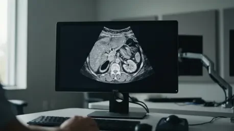

Modern oncology stands at a crossroads where the subtle nuances of tumor biology often escape the limitations of the naked eye, necessitating a shift toward more sophisticated diagnostic frameworks. Evidence suggests that radiomic models consistently outperform traditional clinical models across nearly all types of gastrointestinal malignancies. For decades, medical professionals have relied on the Tumor-Node-Metastasis staging system to dictate treatment paths, yet this method frequently struggles to account for the internal heterogeneity that defines aggressive cancers. Radiomics offers a solution by extracting high-dimensional data from standard imaging like CT, MRI, and PET scans, transforming pixels into actionable biological insights. This computational approach functions as a non-invasive “virtual biopsy,” allowing for the identification of microarchitectural patterns that correlate with patient outcomes. By bridging the gap between qualitative radiology and quantitative data science, clinicians are gaining a clearer view of the complex landscape within gastrointestinal tumors. The ability to decode these hidden signals is not merely a technical triumph but a fundamental change in how the medical community approaches prognosis, moving away from generalized staging and toward a truly individualized assessment of cancer behavior and patient risk profiles.
Evolution of Radiomic Research Trends
The trajectory of computational oncology has moved at a remarkable pace, with scientific interest in the field witnessing a significant surge that reached a definitive peak by 2025. This momentum is largely attributed to the convergence of increased computational processing power and the creation of expansive, high-quality institutional imaging repositories that allow for the training of complex models. A bibliometric look at the landscape reveals that the global research effort is concentrated in specific innovation hubs, notably within China, the United States, Italy, and Germany. These nations have spearheaded the development of interdisciplinary frameworks where radiology, oncology, and advanced data science intersect to redefine diagnostic standards. The proliferation of studies in journals like Frontiers in Oncology and Cancers underscores a growing consensus that imaging is no longer just a visual tool but a deep reservoir of quantitative biomarkers. This era of high-throughput data extraction is characterized by a move toward transparency and methodological rigor, ensuring that the insights gained from these digital snapshots are both reproducible and clinically relevant for a global patient population. By the current year, the infrastructure for this research has matured to support global collaborations that transcend individual hospital networks.
To maintain the integrity of these emerging diagnostic tools, researchers have adopted structured methodologies such as the PRISMA-ScR guidelines to map the rapidly expanding literature. By filtering thousands of records down to a core of high-quality primary research, investigators have been able to isolate the specific variables that contribute most to predictive accuracy in gastrointestinal cancers. This systematic approach has revealed that while the field is maturing, it is also diversifying in its application across different organ systems. The focus has moved beyond simple tumor detection to the much more complex task of grading and prognostication, where the goal is to foresee how a malignancy will evolve over time. This evolution in research methodology has been critical in transitioning radiomics from a speculative academic curiosity to a robust candidate for clinical decision-support systems. As the data pools continue to grow from the current year through 2028, the refinement of these models is expected to provide even deeper insights into the subtle biological markers that dictate the success or failure of various therapeutic interventions in the gastrointestinal tract. Furthermore, the shift toward standardized reporting has ensured that findings can be validated across multiple clinical environments, reducing the risk of localized biases.
Measuring Success in Clinical Predictions
One of the most compelling arguments for the adoption of radiomics is its superior performance in predicting treatment response, particularly in the context of neoadjuvant chemoradiotherapy. Traditional assessment methods often require weeks or months to determine if a therapy is working, but radiomic models have demonstrated the ability to detect early physiological changes in tumor tissue long before they are visible to a human radiologist during a standard review. In various studies, these models have achieved gains in the Area Under the Receiver Operating Characteristic curve of up to 0.18 over clinical-only models. This suggests that the spatial intensity and texture features captured by radiomics provide a more sensitive barometer for therapeutic efficacy than conventional metrics. For a patient facing the grueling side effects of chemotherapy, having an early, data-driven indication of whether the treatment is likely to be successful can be life-altering. It allows oncologists to pivot more quickly to alternative strategies if the initial approach proves ineffective, thereby saving precious time and improving the overall quality of care for those dealing with esophageal, gastric, or rectal malignancies.
Beyond the immediate response to therapy, radiomics has shown significant promise in the prediction of early post-therapeutic recurrence and long-term survival metrics. While predicting overall survival remains a complex challenge due to the myriad of external factors involved, radiomics consistently provides a boost in accuracy ranging from 0.05 to 0.16. These gains are not redundant; they are additive, meaning that the computational data complements the existing histopathological information rather than simply repeating it. This multilayered diagnostic approach creates a more comprehensive profile of the disease, effectively filling in the gaps left by traditional biopsies which may only sample a small portion of a heterogeneous tumor. By identifying high-risk patients who are more likely to experience a relapse, healthcare providers can implement more aggressive follow-up schedules and adjuvant therapies. This proactive stance is essential for improving the prognosis of gastrointestinal cancers, which are notoriously difficult to monitor once the primary treatment phase has concluded. The integration of these digital biomarkers into the clinical workflow represents a major step toward a future where every treatment decision is backed by a holistic, data-driven understanding of the tumor’s trajectory.
Identifying Key Imaging Biomarkers
The effectiveness of radiomics hinges on the extraction of specific imaging features that serve as proxies for the underlying tumor biology. Texture analysis has emerged as the most robust category of these biomarkers, utilizing mathematical frameworks like the Gray-Level Co-occurrence Matrix and the Gray-Level Size Zone Matrix to quantify the heterogeneity of voxel intensities. These metrics essentially perform a “virtual biopsy” by assessing the spatial distribution of gray levels within a scan, which often reflects the cellular density and microvascular structure of the malignancy. Tumors with high levels of architectural complexity and irregularity tend to be more aggressive, and radiomics can quantify this chaos with a level of precision that visual inspection cannot match. In addition to texture, shape descriptors such as sphericity and compactness have proven highly effective for identifying structural changes that correlate with lymph node metastasis. These features provide a functional assessment of the tumor’s growth patterns, helping clinicians determine the likelihood of systemic spread and aiding in more accurate staging of the disease across different imaging modalities.
Despite the broad potential of these biomarkers, a noticeable disparity exists in how research is distributed across different anatomical regions within the gastrointestinal tract. Colorectal and gastric cancers have been the focus of over half of the available high-quality literature, with a strong emphasis on using CT and MRI scans to predict pathological responses and local recurrence. In contrast, hepatobiliary and pancreatic cancers remain significantly underrepresented in primary research, despite their high mortality rates and the urgent need for better prognostic tools. This gap in the literature represents a critical area for expansion, as pancreatic cancer in particular is often diagnosed at late stages when surgical options are limited. Applying the same rigorous radiomic pipelines that have succeeded in colorectal studies to these neglected areas could lead to breakthroughs in how we manage some of the most lethal forms of gastrointestinal disease. Addressing this research imbalance is necessary to ensure that the benefits of quantitative imaging are shared across all patient populations, regardless of the specific organ affected by the malignancy. Expanding the scope to include these difficult-to-treat cancers remains a primary objective for researchers heading into the next several years.
Challenges in Standardization and Implementation
For radiomics to transition from a research tool to a standardized clinical practice, the medical community must overcome significant hurdles related to procedural consistency. Currently, a major challenge lies in the heterogeneity of scanning protocols across different healthcare facilities. Factors such as slice thickness, which can vary from 0.5 mm to 5.0 mm, and the timing of contrast agent administration can drastically alter the numerical values of extracted radiomic features. This variability makes it difficult to compare results between hospitals or to apply a predictive model developed at one institution to patients at another. Without standardized imaging parameters, the field risks producing results that are context-dependent rather than universally applicable. Initiatives such as the Image Biomarker Standardization Initiative have provided a framework for defining radiomic features, yet only a minority of current literature explicitly documents adherence to these guidelines. Achieving widespread protocol standardization is a prerequisite for the reliability and reproducibility required in a high-stakes clinical environment where patient lives are on the line.
Another significant bottleneck in the radiomic pipeline is the process of tumor segmentation, which involves precisely outlining the boundaries of a malignancy on a medical scan. Historically, this has been a manual task performed by trained radiologists, a process that is not only labor-intensive but also prone to significant inter-observer variability. Even highly experienced professionals may disagree on the exact margins of a tumor, and these small differences in segmentation can lead to large variations in the resulting radiomic scores. While there is a growing move toward fully automated algorithmic segmentation using artificial intelligence, the majority of research still relies on human intervention. Automated solutions offer the promise of high-speed, consistent contouring, but they require extensive training on diverse datasets to handle the anatomical variations seen in real-world patients. Until segmentation becomes more efficient and objective, the widespread implementation of radiomics in busy diagnostic departments will remain a logistical challenge, as the time required for manual processing often exceeds the capacity of clinical teams. Streamlining this phase through reliable AI-driven automation is essential for the practical scaling of radiomic analysis.
Transitioning to Deep Learning Models
The technological foundation of radiomics is undergoing a profound shift as researchers move away from manually defined “handcrafted” features toward more sophisticated deep learning architectures. Between the previous decade and 2025, the use of convolutional neural networks in radiomic studies more than doubled, reflecting a preference for models that can automatically learn relevant spatial representations directly from raw image data. Unlike traditional radiomics, where a scientist decides which texture or shape metrics to measure, deep learning allows the computer to discover complex patterns that might be too subtle for human definition. This shift has generally led to higher predictive accuracy, particularly in complex tasks like identifying subtle lymph node involvement or predicting the molecular subtypes of gastric tumors. However, this increased power comes with its own set of trade-offs, primarily the issue of algorithmic opacity. These models often operate as “black boxes,” making it difficult for oncologists to understand the underlying biological rationale behind a high-risk prediction, which can hinder clinical trust in the absence of explainable logic.
Accompanying the rise of deep learning is a persistent “validation crisis” that threatens the credibility of high-accuracy reports in the literature. A staggering majority of studies rely solely on internal validation, testing their algorithms on a subset of the same patient population used to build the model. This practice often leads to “overfitting,” where a model performs exceptionally well on its original data but fails when exposed to new patients from different geographic regions or different scanner vendors. Only a small fraction of current research utilizes external validation on independent datasets from separate institutions. For radiomics to be truly useful in the years from 2026 to 2028, models must demonstrate their robustness across diverse clinical settings. Ensuring that an algorithm remains accurate when faced with different patient demographics and varying imaging hardware is the ultimate test of its clinical utility. Without this multi-institutional testing, the impressive results seen in experimental settings may prove to be overly optimistic best-case scenarios that do not translate to better patient outcomes in the real world. Addressing this lack of external verification is a top priority for the ongoing maturation of the field.
Moving Toward Clinical Integration
To bridge the final gap between laboratory research and the patient’s bedside, the focus of the medical community shifted toward the development of explainable artificial intelligence. Clinicians required more than just a probability score; they needed to understand which biological characteristics of the tumor, such as cellular density or vascular heterogeneity, were driving the model’s conclusions. By prioritizing explainability, developers created tools that not only predicted outcomes but also provided a deeper understanding of the disease itself, fostering a stronger sense of trust among the oncology teams. Furthermore, the field advanced through the initiation of large-scale, prospective randomized trials. While retrospective studies provided a strong proof of concept, prospective data served as the gold standard required to prove that radiomics-guided treatment strategies led to improved survival rates and a better quality of life for patients. These rigorous real-world tests successfully validated radiomics as a cornerstone of personalized gastrointestinal oncology, setting a new benchmark for evidence-based medicine during the current year.
The path forward involved a fundamental shift in the relationship between human expertise and machine intelligence, which moved toward a model of collaborative decision-support. Rather than viewing radiomics as a replacement for the radiologist, the medical community successfully positioned these tools as sophisticated assistants that handled high-volume data processing while human experts provided essential clinical context. This partnership allowed for the identification of scanning artifacts and anatomical variations that computers occasionally misinterpreted, ensuring a higher level of diagnostic safety. Efforts to standardize imaging protocols and adopt unified reporting guidelines bore fruit, creating a more cohesive environment for data exchange across international borders. The integration of radiomic scores into the broader clinical picture, alongside genomic and proteomic data, marked the beginning of a truly multi-omic approach to cancer care. These actionable steps transformed radiomics from a high-tech experimental niche into a practical, data-driven framework that significantly enhanced the precision of treatment for gastrointestinal malignancies, ensuring that clinical decisions were more robust than ever before.