
The emergence of compressed-sensing MP2RAGE technology has reduced MRI acquisition times to under four minutes while maintaining the high resolution necessary for tissue characterization. This breakthrough represents a pivotal moment in neuroimaging, as it addresses the long-standing need for a noninvasive and cost-effective method to track the molecular indicators of Alzheimer’s disease. For decades, the presence of amyloid-beta plaques has been the definitive marker for the condition, yet detecting these clusters typically involved invasive cerebrospinal fluid analysis or expensive positron emission tomography scans. Recent research indicates that measuring T1 relaxation times—how long it takes for protons to return to equilibrium after a radiofrequency pulse—serves as a robust indicator of the brain’s internal environment. By utilizing quantitative mapping, clinicians can now look beyond simple brain volume loss to identify the subtle microstructural shifts that signal the early onset of neurodegenerative pathology in patients. This advancement is vital as the global medical community shifts toward earlier detection and personalized intervention strategies for neurodegeneration in 2026.
Technological Advancements in Neuroimaging
Reducing Acquisition Barriers via Compressed-Sensing
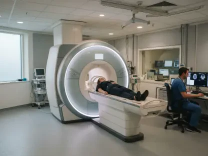
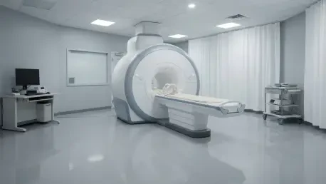
A primary challenge in implementing quantitative magnetic resonance imaging within a clinical setting has historically been the lengthy acquisition times required for high-quality data. Standard protocols often demand over eight minutes of patient immobility, a requirement that is frequently impossible for individuals suffering from cognitive impairment or physical restlessness associated with neurodegeneration. To overcome this, the study utilized a 3-tesla MRI scanner equipped with an advanced compressed-sensing MP2RAGE sequence, which effectively slashed the necessary scan time to approximately three minutes and forty seconds. This efficiency is not merely a convenience; it is a critical evolution that allows for the practical integration of complex biomarkers into the daily routine of busy hospitals and diagnostic centers. By significantly reducing the window for potential patient movement, the technology ensures that the resulting data remains sharp and clinically actionable for further analysis.
This specific technical leap is significant because it maintains high-resolution, three-dimensional images while being fast enough for routine clinical use. It allows for the simultaneous collection of structural data and tissue characterization, making the diagnostic process far more efficient for both doctors and patients. In 2026, the speed of this imaging protocol does not sacrifice the quality of the data, ensuring that patients who may struggle with cognitive distress can still undergo complete evaluations without the need for sedation or multiple sessions. By making high-quality quantitative data accessible in such a short timeframe, the research paves the way for T1 mapping to move from an experimental tool to a practical diagnostic staple. This efficiency also reduces the overall cost of imaging, as more patients can be scanned within a standard workday, effectively lowering the barrier to entry for advanced neurological screenings in various medical facilities across the country.
Balancing Speed with High-Resolution Data
Despite the dramatic increase in imaging speed, the compressed-sensing technique does not compromise the spatial precision required for neuroanatomical evaluation. The protocol maintains a 1-millimeter isotropic resolution, delivering a high-definition view of the brain’s architecture that is essential for detecting the minute alterations associated with early-stage Alzheimer’s. This balance of speed and precision allows for the simultaneous acquisition of three-dimensional T1-weighted structural images and quantitative maps, providing a comprehensive assessment of both tissue volume and composition in a single scan. Such a multifaceted approach enables medical professionals to streamline the diagnostic workflow, moving away from fragmented testing toward a more unified and patient-friendly evaluation process. The ability to generate these detailed maps within a standard clinical appointment timeframe marks a significant departure from previous research, positioning T1 mapping as a viable tool for widespread medical use.
Furthermore, the implementation of this high-resolution mapping facilitates a more nuanced understanding of the brain’s microstructural integrity. By providing a quantitative measure that goes beyond simple volume loss, T1 mapping offers a window into the actual health of the tissue remaining. This is particularly important for differentiating between normal age-related changes and the early pathological shifts of Alzheimer’s disease. In 2026, clinicians are increasingly relying on these quantitative metrics to provide a more objective basis for diagnosis, reducing the reliance on subjective interpretations of visual scans. The precision afforded by the 1-millimeter isotropic resolution ensures that even the smallest regions of interest, such as specific subfields of the temporal lobe, can be analyzed with confidence. This level of detail is necessary for the development of targeted therapies that aim to preserve brain function at the earliest possible stage of the neurodegenerative process.
Regional Analysis and Disease Validation
Identifying Alterations in the Hippocampus and Cortex
The central focus of this imaging research centered on the hippocampus, an area of the brain deeply involved in memory formation and typically the first to experience the impact of Alzheimer’s. The data revealed that individuals diagnosed with mild cognitive impairment exhibited significantly higher T1 relaxation times, averaging 1,398 milliseconds, compared to 1,349 milliseconds in the healthy control group. This statistically significant difference underscores the sensitivity of T1 mapping to the microscopic changes occurring within the hippocampal tissue. Curiously, when the researchers examined these values alongside cognitive test scores, they found no direct linear correlation between the T1 measurement and the severity of clinical symptoms. This finding is particularly insightful, as it suggests that T1 relaxation changes might emerge as a very early biological signal, potentially appearing before a patient begins to manifest the clear cognitive decline traditionally used for clinical diagnosis.
In addition to the hippocampal findings, a voxel-wise analysis provided a more granular look at how T1 values shift throughout the brain’s cortices. The researchers identified focal increases in relaxation times within the temporal, parietal, and occipital regions, areas often associated with the later spread of neurodegeneration. A noteworthy observation was that these T1 alterations were more localized and specific than the general cortical atrophy that typically characterizes advanced stages of the disease. This implies that T1 mapping captures distinct microstructural transformations—likely related to tissue composition and health—that occur independently of, or even prior to, the physical shrinking of brain matter. By pinpointing these localized changes, clinicians may be able to identify the unique signature of Alzheimer’s disease long before widespread volume loss becomes apparent on a standard scan, allowing for much earlier clinical intervention and monitoring in 2026.
Monitoring Pathology through Quantitative Mapping
To validate the biological accuracy of these MRI findings, the research team compared the T1 maps against amyloid-PET scans, which are widely considered the gold standard for measuring protein burden in the brain. The results demonstrated a robust positive association between T1 relaxation times and the standardized uptake value ratios from the PET data, confirming that as T1 values increased, so did the density of amyloid-beta plaques. Although the observed effect size was modest, the high level of statistical significance proves that a rapid, noninvasive MRI sequence can effectively mirror the information provided by more invasive or radioactive molecular imaging techniques. This correlation is essential for establishing T1 mapping as a credible biomarker, as it provides a direct link between the physics of magnetic resonance and the underlying molecular pathology that drives the progression of Alzheimer’s disease in a living patient.
The biological mechanisms driving these elevated T1 values were rooted in the physical environment created by plaque accumulation. As amyloid-beta deposits built up in the brain, they tended to expand the extracellular space, which in turn led to an increase in local water content. Since water molecules have naturally long T1 relaxation times, their increased presence within the brain tissue directly inflated the overall T1 measurement. Furthermore, the degradation of myelin—the protective insulation surrounding nerve fibers—was known to occur alongside amyloid deposition and also contributed to the prolongation of these relaxation times. Longitudinal studies were initiated to determine if these T1 shifts could track the real-time efficacy of new amyloid-clearing therapies, which was a vital step for drug development in 2026. This approach enabled a shift toward more personalized medicine, where treatment adjustments were made based on rapid, noninvasive imaging feedback. Ultimately, the integration of compressed-sensing MRI into standard neurological care provided a more accessible pathway for identifying the silent stages of neurodegeneration.