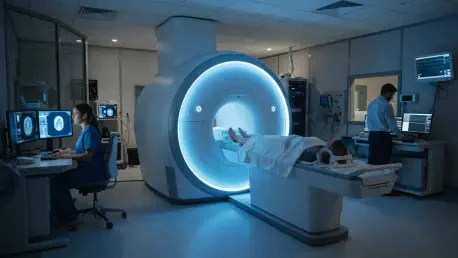

While 6Ga-PSMA and 18F-DCFPyL are cornerstones of modern prostate cancer diagnostics, their effectiveness is limited by the respiratory ‘blur’ that occurs in the chest and abdomen. This phenomenon is a persistent technical obstacle in nuclear medicine because Positron Emission Tomography/Computed Tomography scans require several minutes of data acquisition, during which patients must breathe naturally. The constant movement of the diaphragm creates motion artifacts, particularly in the lower thorax and upper abdomen, causing small metastatic lesions to appear more diffuse and less intense than they are in reality. This lack of clarity often complicates the staging of prostate cancer, potentially leading to the underestimation of a patient’s total disease burden. To address this, a recent retrospective study published in EJNMMI Reports evaluated the potential of data-driven gating. By using the OncoFreeze AI algorithm, researchers sought to determine if software-based motion correction could enhance image quality without the need for external hardware.
The Evolution: PSMA Tracers and Gating Technology
The effectiveness of PSMA-targeted imaging is largely determined by the specific radiopharmaceutical employed, with Gallium-68 and Fluorine-18 being the primary agents in clinical use today. While both are exceptional at identifying cancerous cells, they possess distinct physical properties, such as different positron ranges, which can affect the ultimate resolution of the scan. In the context of the growing theranostics field, where diagnostic scans dictate a patient’s suitability for radioligand therapy, ensuring the highest possible image quality is a clinical necessity. If a tracer like 68Ga-PSMA, which is often paired with therapeutic 177Lu-PSMA-617, produces images affected by motion blur, clinicians may struggle to identify low-grade metastases or distant lymph node involvement. Consequently, optimizing these scans has become a primary objective for oncology departments seeking to refine their diagnostic accuracy and ensure that patients receive the most appropriate treatments based on their specific disease profile.
Historically, clinicians attempted to mitigate the effects of respiratory motion through mechanical means, such as external pressure-sensitive belts or respiratory bellows. These devices track the breathing cycle and allow the scanner to “gate” the data, but they are frequently cumbersome to use and prone to technical failure during busy clinical hours. Furthermore, traditional gating methods often lead to significantly increased image noise because they discard a large portion of the detected photon counts—sometimes up to 80%—to isolate a single phase of the respiratory cycle. Modern data-driven gating techniques like OncoFreeze AI represent a significant departure from these limitations by extracting motion information directly from the PET “list-mode” data. Because the software identifies breathing patterns mathematically from the signal itself and utilizes the entire set of acquired data during reconstruction, it manages to maintain high signal-to-noise ratios while simultaneously sharpening the resulting images.
Quantitative Improvements: Enhancing Metastatic Detection
In a comprehensive analysis involving a cohort of 63 patients, researchers identified 325 distinct metastatic lesions to evaluate the performance of data-driven gating across various anatomical regions. These lesions included bone metastases, lymph node involvements, and lung nodules, providing a robust dataset for comparing standard static reconstructions against the newer AI-corrected versions. The primary metric used to assess these improvements was the Standardized Uptake Value, or SUV, which serves as the industry standard for quantifying the metabolic activity of a tumor. The findings demonstrated a consistent and statistically significant rise in SUV measurements when motion correction was applied. This increase is critical because it indicates that the scanner is capturing a more concentrated and accurate representation of the tracer within the lesion. By correcting for the physical displacement caused by breathing, the algorithm effectively “re-focuses” the signal that would otherwise be spread across several millimeters.
The most dramatic gains in quantitative accuracy were observed in the lung lesions, where physical displacement during the breathing cycle is most extreme. For patients injected with 18F-PSMA, the SUV measurements in pulmonary metastases increased by over 32% following the application of the gating software, while the 68Ga-PSMA group experienced a 14.4% boost in the same category. These results highlight the specific vulnerability of thoracic imaging to motion-induced errors and underscore the value of software-based stabilization. Even in bone and lymph node lesions, which are generally more stable, the improvements in contrast-to-noise ratios were substantial. For instance, lymph node detection in the 68Ga-PSMA cohort saw a 21.7% improvement in contrast, allowing small, high-risk nodes to stand out more clearly against the surrounding physiological background. Such improvements ensure that radiologists can differentiate between true disease and incidental uptake or noise, which is essential for accurate cancer staging and monitoring.
Visual Clarity: Radiologist Consensus and Sharpness
Beyond the objective numerical data, the clinical utility of any imaging technology depends heavily on the subjective interpretation of the medical professional. To test this, two experienced radiologists conducted a blinded review of the datasets, comparing traditional static images against those processed with data-driven gating. Their qualitative assessment focused on parameters such as overall image quality, tumor sharpness, and the presence of artifacts that might interfere with a diagnosis. The results were remarkably consistent, with the gated images receiving a “high” quality rating in 95% of the evaluations. Furthermore, in 97% of the cases, the clinicians determined that the gated reconstructions provided superior tumor sharpness. This visual enhancement is particularly important when evaluating the margins of a lesion or its proximity to critical anatomical structures. When images are crisper, the diagnostic confidence of the physician increases, reducing the likelihood of ambiguous findings.
The shift toward software-driven solutions also offers significant operational advantages within a high-volume clinical environment. By eliminating the need for external belts and sensors, oncology departments can streamline their imaging workflows, reducing the time required to set up each patient and minimizing the risk of hardware-related data loss. OncoFreeze AI functions as a background process that utilizes the raw list-mode data already being captured by the PET scanner, making it a seamless addition to existing protocols. This efficiency is paired with a noticeable reduction in visual noise, which has historically been a major complaint regarding gated imaging. By using the full count of detected photons rather than a small fraction, the AI-driven approach provides a smooth, clear background that makes small, bright spots of metastatic activity much easier to identify. This balance between noise reduction and signal enhancement represents the current state of the art in molecular imaging, providing a tool that is technically superior.
Clinical Impact: The Path to Precision Theranostics
The integration of data-driven gating is particularly vital in the context of theranostics, where accurate diagnostic measurements directly influence life-altering treatment decisions. For example, current clinical guidelines suggest that patients with an average SUV above a certain threshold on a PSMA PET scan are the most likely to respond favorably to targeted therapies like 177Lu-PSMA-617. If respiratory motion artificially lowers these measured values, a patient might be incorrectly excluded from a treatment that could significantly extend their life. By providing a “true” SUV that reflects the actual concentration of the radiopharmaceutical in the tumor, data-driven gating acts as a safeguard for patient selection. It ensures that the criteria used to qualify patients for advanced therapies are based on the most precise data possible, rather than being distorted by the physiological movements of the patient’s body during the scanning process, thereby maximizing the potential for successful clinical outcomes.
Furthermore, these advancements align with broader global research trends that favor hardware-free motion correction across all types of nuclear medicine. While earlier studies focused on general oncology tracers like 18F-FDG, the current validation of these techniques for PSMA-specific agents is a crucial milestone for urological oncology. Every tracer has a unique biodistribution and signal profile, meaning that software must be specifically calibrated to handle the nuances of prostate cancer imaging. The success of the OncoFreeze AI algorithm in this specific patient population demonstrates that software-based motion correction is a versatile and robust solution that can be adapted to various clinical needs. As medical centers around the world continue to adopt these technologies, the standardization of image quality across different scanner models and institutions becomes a more achievable goal. This movement toward consistency is essential for conducting large-scale multi-center clinical trials, where uniform data collection is necessary.
Future Integration: Addressing Technical Refinements
Despite the significant progress documented in recent years, technical challenges remain that provide a roadmap for future developmental efforts in medical imaging. One notable issue is the “misregistration” that can occur between motion-corrected PET data and the corresponding CT scan, which is typically acquired as a static snapshot. Because the PET signal is being adjusted to account for breathing but the CT map remains uncorrected, the two layers of information may not perfectly overlap. This misalignment can lead to inaccuracies in both anatomical localization and the attenuation correction process, which is why future research is currently focusing on the integration of 4D-CT. By syncing the motion correction across both the PET and CT components of the scan, developers aim to achieve a state of perfect anatomical registration. Such a breakthrough would further enhance the precision of radiotherapy planning and surgical interventions, where even a few millimeters of error can have significant consequences.
To fully realize the benefits of data-driven gating, healthcare providers should begin integrating these software protocols into their standard diagnostic workflows. The transition from retrospective research to prospective clinical application was historically slow, but the current accessibility of AI-driven tools has accelerated this timeline significantly. Moving forward, it is recommended that oncology centers prioritize the training of radiological staff in interpreting gated images, as the increased sharpness and SUV values may require adjustments to traditional reading thresholds. The study successfully demonstrated that software-based solutions could solve long-standing hardware limitations, paving the way for a new era of high-definition molecular imaging. By adopting these methods, the medical community took a decisive step toward more reliable and personalized cancer care. Future efforts must continue to expand the use of these algorithms to other motion-prone areas, such as the liver and heart.