
Modern silicon photomultiplier detectors offer superior photon sensitivity and spatial resolution compared to the traditional photomultiplier tubes used in older imaging equipment. This technological leap has arrived at a critical juncture in the fight against neurodegenerative diseases, where the precise visualization of tau protein serves as a definitive roadmap for cognitive decline. Unlike amyloid beta plaques, which often appear years before symptoms manifest and correlate poorly with the severity of impairment, tau aggregates provide a direct look at the active destruction of neuronal architecture. Encoded by the microtubule-associated protein tau gene, these filaments disrupt cellular stability and spread in a predictable hierarchical fashion across the brain. By 2026, the ability to accurately stage this progression through positron emission tomography has become a cornerstone of dementia care, allowing clinicians to distinguish between normal age-related changes and more aggressive pathologies such as Alzheimer’s disease or progressive supranuclear palsy. However, the success of these molecular insights historically depended on the patient’s ability to remain perfectly still during lengthy scans, creating a significant barrier for those in the advanced stages of cognitive loss.
The Technological Evolution: Radiotracers and Imaging Hardware
The introduction of second-generation radiotracers has fundamentally redefined what is possible in the realm of neuroimaging, with fluorine-18-MK6240 leading the charge toward higher diagnostic clarity. Unlike earlier iterations of tau probes that were often plagued by off-target binding to monoamine oxidase in the basal ganglia and other brain regions, MK6240 offers an exceptionally clean signal with a high binding affinity for tau filaments. This specific tracer allows for the clear visualization of the disease as it moves from the entorhinal cortex into the limbic system and eventually the association cortices. The high signal-to-noise ratio inherent in this tracer means that the molecular signature of the disease is more pronounced, which naturally raises the question of whether the extensive acquisition times utilized in older protocols are still necessary. In the current clinical environment, where accuracy and speed must coexist, these chemical advancements provide the raw data required to explore significantly shorter scanning windows without sacrificing the integrity of the results.
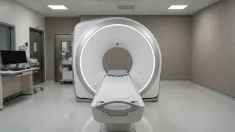
Parallel to the development of these advanced tracers is the widespread adoption of high-sensitivity imaging hardware that utilizes silicon photomultiplier detectors and time-of-flight technology. These systems are designed to detect even the faintest photon signals and accurately localize annihilation events by measuring the infinitesimal time differences between photon detections. By maximizing the efficiency of every radioactive decay event, these scanners can generate high-resolution images even when the total number of detected photons is lower than what was traditionally required. This synergy between advanced chemical probes and state-of-the-art detector technology suggests that the standard twenty-minute scan may be an outdated benchmark. If a scanner can collect a sufficient density of data in just five minutes to determine a patient’s tau status, the implications for patient comfort and clinic efficiency would be transformative, particularly for elderly populations who struggle with the physical demands of staying motionless in a narrow scanner bore for extended periods.
Clinical Methodology: Reconstructing the Acquisition Window
A comprehensive retrospective study conducted at a prominent memory clinic between late 2024 and early 2025 aimed to rigorously validate the potential of these shortened scan times. The investigation included sixty-nine patients, with a demographic split of 42 women and 27 men, all of whom presented with various stages of mild cognitive impairment or Alzheimer’s disease. Every participant underwent a specialized imaging protocol that began with an intravenous injection of the 18F-MK6240 tracer, followed by a controlled ninety-minute uptake period to ensure the tracer had fully bound to the target proteins in the brain. The scans were performed on an integrated 3T GE Healthcare SIGNA PET/MR system, which allowed the team to capture high-resolution anatomical structures and molecular data simultaneously. This simultaneous acquisition was vital for creating a precise anatomical roadmap that ensured the molecular signals were correctly assigned to specific regions of interest within the brain’s complex architecture.
The researchers employed a sophisticated “list-mode” data acquisition strategy, which essentially records every individual photon detection as a discrete event with a specific timestamp. This raw format allowed the scientists to retrospectively reconstruct the images into four distinct time durations: five, ten, fifteen, and twenty minutes. By using this method, the team could compare different acquisition lengths for the exact same patient session, eliminating the variables that would arise if the patient had been scanned multiple times. These reconstructions were then subjected to rigorous qualitative and quantitative analysis to determine if the five-minute snapshot could replicate the diagnostic power of the twenty-minute gold standard. The methodology focused heavily on regional brain zones, specifically examining the sixteen areas associated with the Braak staging system, which is the international standard for describing the anatomical spread of tau pathology and its corresponding severity in the human brain.
Diagnostic Performance: Visual and Quantitative Alignment
The qualitative phase of the research involved a blinded review by senior neuroimaging specialists who assessed the images for noise levels, overall clarity, and diagnostic confidence. The results provided a compelling case for shorter protocols, as every five-minute reconstruction was rated as being of adequate or excellent diagnostic quality. While two-minute reconstructions were often discarded due to excessive noise that obscured fine details, the five-minute scans maintained enough structural and molecular signal to allow for definitive interpretations. Crucially, when the specialists were tasked with providing a binary diagnosis of tau-positivity, there was total agreement between the five-minute and twenty-minute results. This suggests that the fundamental question of whether a patient has a pathological tau burden can be answered with absolute certainty in just a fraction of the traditional time, effectively eliminating the need for prolonged patient exposure to the imaging environment.
Moving beyond subjective visual assessments, the quantitative data further solidified the reliability of the truncated scan window. The researchers calculated standardized uptake value ratios by comparing the tracer signal in target brain regions to the signal in the cerebellar gray matter, which serves as a stable reference point. The statistical analysis showed that the mean ratios remained remarkably consistent across all acquisition durations, with no significant deviations observed between the five-minute and twenty-minute data blocks. Receiver operating characteristic curve analysis revealed that the discriminative power of the scan—its ability to distinguish between those with and without disease—stayed nearly identical, with an area under the curve of 0.982 for the five-minute window. Furthermore, the inter-reader agreement for regional Braak staging was nearly perfect, indicating that the shortened scan does not lose the ability to track how far the disease has spread throughout the different lobes of the brain.
Strategic Implications: Efficiency and Future Research Standards
The transition to a five-minute PET scan protocol offers a multi-layered set of benefits that extend from the patient experience to the operational logistics of the healthcare system. From a patient-centric perspective, reducing the time spent in the scanner by 75 percent drastically lowers the probability of motion-induced artifacts, which are the primary cause of poor image quality in dementia imaging. Shorter scans reduce the physical and psychological burden on patients, many of whom may suffer from claustrophobia or the agitation common in memory disorders. For clinical centers, this shift allows for a significant increase in patient throughput, enabling facilities to provide life-changing diagnoses to a larger portion of the aging population. By optimizing the use of expensive PET/MR equipment, hospitals can reduce wait times and improve the overall return on investment for their advanced imaging suites, all while maintaining the highest possible standards of diagnostic accuracy.
Despite the clear successes of the five-minute protocol for routine clinical use, the study highlighted specific scenarios where the standard twenty-minute scan remains the necessary benchmark. The researchers observed that while the average values remained stable, individual measurements became slightly more variable at the five-minute mark due to the naturally lower photon statistics. This variance is negligible for a one-time diagnostic assessment, but it could be problematic in longitudinal clinical trials where researchers are looking for extremely small, incremental changes in tau levels to measure the effectiveness of a new drug. Consequently, a tiered approach was proposed: utilizing the ultra-fast five-minute scan for frontline clinical diagnosis and reserving the twenty-minute scan for high-precision research and long-term monitoring. Future multi-center trials were identified as the next logical step to ensure these findings remain consistent across different scanner brands and include more borderline cases where tau levels are at the very edge of detection.