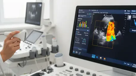

Finding a definitive biological marker for Parkinson’s disease has historically required a combination of invasive procedures and prohibitively expensive neuroimaging that remains inaccessible for many patients worldwide. While magnetic resonance imaging and positron emission tomography provide detailed views of the brain, their high costs and the need for specialized facilities create significant barriers to early diagnosis. Parkinson’s disease is characterized by the progressive loss of dopaminergic neurons, yet identifying this decline in its nascent stages is notoriously difficult using standard clinical examinations. Traditional diagnostic pathways often rely on the subjective observation of motor symptoms, which may only appear after significant neurological damage has already occurred. To address these limitations, researchers have developed a sophisticated computational framework that utilizes stacked artificial intelligence models to analyze brain ultrasound data. This innovation represents a major step forward in creating a reliable, low-cost diagnostic tool.
Standardizing Ultrasound Interpretation: The Shift to Algorithmic Diagnostics
Transcranial sonography has long been recognized as a safe and relatively inexpensive method for visualizing the substantia nigra, a brain region central to the development of Parkinson’s disease. In patients with the condition, this area often exhibits a phenomenon known as hyperechogenicity, appearing as unusually bright spots on an ultrasound scan due to changes in tissue composition. Despite these advantages, the clinical utility of ultrasound has been hindered by a high degree of inter-observer variability, where different physicians might interpret the same image differently. The quality of these scans is also highly dependent on the operator’s skill and the patient’s physiological characteristics, such as the thickness of the temporal bone window. These inconsistencies have traditionally relegated ultrasound to a secondary role in the diagnostic process rather than a primary tool. However, the introduction of automated analysis aims to standardize these interpretations across the medical field.
The implementation of an artificial intelligence framework effectively eliminates the guesswork associated with manual image review by applying consistent mathematical criteria to every scan. By utilizing advanced algorithms to detect patterns of hyperechogenicity, the system can identify markers of disease with a level of precision that exceeds the capabilities of the human eye. This shift from qualitative observation to quantitative analysis ensures that diagnostic results remain stable regardless of the equipment used or the experience level of the technician. Furthermore, the model is designed to account for technical limitations, such as low-contrast images or noise, which frequently occur in real-world clinical settings. By providing a standardized baseline for evaluation, the technology allows for more reliable longitudinal tracking of a patient’s condition. This objectivity is essential for developing large-scale screening programs that can accurately identify at-risk individuals before their motor functions begin to deteriorate significantly.
Hierarchical Intelligence: How Stacked Models Improve Accuracy
The core of this diagnostic advancement lies in its stacked machine learning architecture, which functions by organizing various algorithms into a hierarchical structure to maximize predictive power. In the initial stage of this process, several independent base models are trained to analyze raw data from ultrasound scans through different lenses of interpretation. Some of these models are specifically optimized to detect subtle variations in the texture of brain tissue, while others focus on the volumetric properties and spatial boundaries of the substantia nigra. This diversified approach ensures that no single feature is overlooked, as the system captures a wide range of morphological characteristics simultaneously. By leveraging the strengths of multiple specialized algorithms, the framework avoids the common pitfalls of single-layer models that might be overly sensitive to one specific type of data artifact. This collaborative processing of information creates a robust foundation for the final diagnostic assessment.
Once the base models have completed their respective analyses, a secondary meta-model synthesizes their individual findings into a single, cohesive diagnostic output. This meta-model is trained to recognize which base algorithms perform most reliably under specific conditions, effectively weighting their contributions based on historical accuracy and data quality. By examining the correlations between the outputs of the various base layers, the system can filter out contradictory signals and highlight the most consistent indicators of neurological decline. This hierarchical approach, known as ensemble learning, significantly reduces the likelihood of false positives and negatives compared to conventional artificial intelligence methods. The resulting diagnostic precision allows clinicians to make more informed decisions with higher confidence in the underlying data. This structural complexity is what enables the system to maintain high performance across diverse patient populations, making it a versatile tool for various clinical environments.
Multi-Modal Fusion: Combining Imaging and Clinical Metrics
To further enhance the accuracy of the diagnostic process, the researchers incorporated a technique called multi-modal fusion, which integrates ultrasound image data with diverse clinical information. This holistic approach recognizes that Parkinson’s disease is not merely a structural condition but one that manifests through a variety of behavioral and demographic factors. The AI system is designed to ingest non-imaging data such as patient age, gender, and the results of standardized motor skill assessments, blending these variables with the visual findings from the scans. By contextualizing the ultrasound data within a broader clinical framework, the model can differentiate between age-related brain changes and the specific pathological markers of Parkinson’s. This integration is particularly valuable for identifying early-stage patients who may show only subtle imaging abnormalities but demonstrate more pronounced clinical signs. The fusion of these data streams creates a comprehensive profile for each individual.
The synergy achieved through multi-modal fusion allows the artificial intelligence to uncover complex correlations that might remain invisible during a standard medical review. For instance, the system may identify that a specific degree of tissue hyperechogenicity is significantly more predictive of disease when paired with certain tremor characteristics or cognitive metrics. These hidden relationships provide a more nuanced understanding of the disease’s heterogeneity, allowing for more personalized diagnostic assessments. This capability is vital because Parkinson’s disease often presents differently across various demographics, making a “one-size-fits-all” approach to imaging less effective. By considering the patient as a whole rather than a set of isolated symptoms, the AI provides a more accurate reflection of the underlying pathology. This comprehensive analysis not only improves the initial diagnosis but also helps in categorizing different subtypes of the disease, which is essential for tailoring future treatment strategies.
Explainable AI: Building Trust Through Interpretability and Transparency
A significant challenge in the adoption of artificial intelligence within the medical field has been the “black-box” nature of complex algorithms, where the reasoning behind a decision is unclear. To overcome this barrier, the researchers prioritized the development of explainable AI tools that provide transparency into how the model reaches its conclusions. These features allow clinicians to see exactly which areas of an ultrasound scan or which clinical data points were most influential in generating a particular diagnosis. By visualizing the decision-making process through heat maps and feature-importance charts, the technology acts as a collaborative assistant rather than a mysterious substitute for medical expertise. This transparency is crucial for building trust between healthcare providers and automated systems, ensuring that AI recommendations are always verifiable through human clinical judgment. When doctors can understand the logic behind a diagnosis, they are better equipped to explain the results to their patients.
The portability and cost-effectiveness of ultrasound technology, combined with the power of stacked AI models, offer a transformative solution for improving diagnostic access in underserved regions. Unlike expensive imaging equipment that requires specialized infrastructure, portable ultrasound machines can be deployed in rural clinics and community health centers. By integrating advanced AI directly into these accessible platforms, high-quality neurological diagnostics can be delivered to populations that previously lacked access to specialized care. This democratization of technology has the potential to facilitate earlier interventions on a global scale, leading to better management of the disease and improved quality of life for millions of people. Furthermore, the underlying logic of this stacked framework is adaptable, suggesting that similar models could eventually be trained to detect other neurodegenerative conditions. This scalability ensures that the benefits of this research extend far beyond a single disease or geographic location.
Practical Implementation: Redefining Global Neurological Care Standards
The integration of stacked machine learning into clinical workflows fundamentally changed the approach to diagnosing chronic neurological conditions within the healthcare system. Medical centers that adopted these automated sonography tools reported a significant decrease in the time required to reach a definitive diagnosis for symptomatic patients. By utilizing the objective data provided by the meta-modeling framework, hospitals successfully reduced the rate of misdiagnosis that previously plagued traditional assessment methods. Researchers observed that the early identification of substantia nigra changes allowed for the initiation of neuroprotective therapies much sooner than in previous years. This proactive stance led to improved long-term outcomes for patients, as early intervention helped to mitigate the severity of motor symptoms over time. Public health organizations emphasized the importance of training local staff to operate the portable equipment while relying on the AI to provide high-level analytical support for those in remote areas.
Following the success of these Parkinson’s diagnostic protocols, the medical community took strategic steps to expand the application of stacked AI models to other neurodegenerative disorders. Engineers worked closely with neurologists to adapt the multi-modal fusion techniques for identifying early markers of dementia and multiple sclerosis. This shift toward a more comprehensive digital health infrastructure enabled a more efficient allocation of resources, focusing expensive neuroimaging only on the most complex cases identified by the initial AI screening. Healthcare providers also focused on integrating these tools into electronic health records to ensure that diagnostic data remained accessible across different levels of care. These actions established a new standard for precision medicine that prioritized data transparency and patient accessibility. By moving toward a more collaborative relationship between human expertise and algorithmic precision, the industry successfully created a more resilient and equitable framework for neurological health.