
The long-standing dominance of Next-Generation Sequencing (NGS) as the gold standard for identifying genetic mutations in cancer is facing its first major structural challenge from the field of computer vision. For decades, oncologists have relied on the extraction and chemical analysis of genetic material to understand the underlying drivers of a patient’s malignancy, a process that is famously resource-intensive and slow. However, recent breakthroughs in deep learning and image analysis have demonstrated that the morphology of cells—the way they look under a standard microscope—contains hidden signatures that correlate directly with complex genomic alterations. By leveraging these visual patterns, a new generation of diagnostic platforms is beginning to offer a path toward rapid, low-cost screening that could fundamentally change how laboratories prioritize and treat various forms of cancer.
Shifting Paradigms in Precision Oncology
Bridging the Gap Between Morphology and Genomics
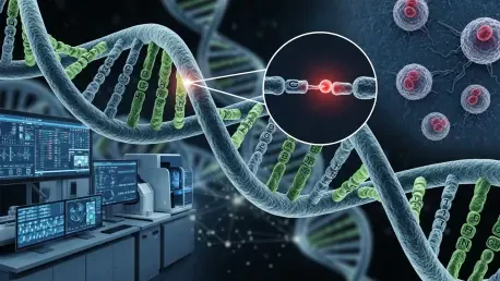
The traditional diagnostic pipeline often begins with a biopsy and a routine stained slide, which is then followed by a separate, expensive journey into the world of molecular genetics. This disconnect between what a pathologist sees and what a sequencer detects has long been considered an unavoidable necessity of modern medicine. However, the emergence of advanced AI platforms like those developed by Moonlight AI AG is proving that these two worlds are far more interconnected than previously thought. By analyzing digital scans of standard blood and cytology smears, specialized algorithms can now detect the subtle architectural changes in cells that indicate specific genomic biomarkers. This capability effectively turns a simple image into a molecular map, allowing clinicians to gain high-level genetic insights without immediately resorting to the destructive and time-consuming process of chemical DNA extraction.
This shift toward image-based genomics represents a major leap in how diagnostic data is harvested from existing clinical samples. Instead of treating the physical slide as merely a visual reference for a human pathologist, technology now treats every pixel as a data point that can be cross-referenced against massive genomic databases. By identifying disease signatures directly from routine imaging, laboratories can significantly reduce their overhead while maintaining a high degree of diagnostic accuracy. This approach does not necessarily aim to eliminate DNA sequencing entirely but rather to act as a sophisticated filter that provides immediate answers for common mutations. Consequently, the reliance on NGS is transitioning from a mandatory first step to a targeted secondary confirmation, streamlining the entire diagnostic workflow for hematology and oncology departments.
Scaling Diagnostic Capacity Through Advanced Software
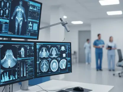
The logistical hurdles of implementing high-end genomic testing are often prohibitive for smaller clinical settings or those in developing healthcare markets where hardware investment is limited. Integrating AI-powered diagnostic software allows these facilities to leverage their existing infrastructure—such as standard digital scanners and microscopes—to perform tasks that previously required a specialized molecular laboratory. Because these software solutions do not require new hardware or manual interventions, they can be scaled rapidly across a network of hospitals with minimal friction. This democratization of high-level diagnostics ensures that a patient’s access to precision medicine is no longer strictly determined by the physical location or the budget of the clinic they happen to visit.
Furthermore, the automation of image analysis addresses the critical shortage of specialized hematopathologists and cytotechnologists by handling the repetitive and labor-intensive aspects of slide review. By pre-screening samples and flagging those with high probabilities of specific genetic markers, the AI serves as an intelligent triage system. This allows human experts to focus their attention on complex or borderline cases where professional judgment is most vital. The efficiency gains are not merely administrative; they translate directly into faster turnaround times for patients who are waiting for life-altering treatment decisions. As these platforms continue to evolve through 2026 and beyond, the capacity for laboratories to process thousands of samples with high precision becomes a reality rather than a theoretical ambition.
Strategic Implementation and Global Integration
Building Robust Datasets for Clinical Reliability
For any AI-driven diagnostic tool to gain widespread acceptance in the medical community, it must be backed by a diverse and massive foundation of verified data. Moonlight AI has recognized this requirement by establishing an international consortium of clinical partners to build a first-of-its-kind cytopathology database. This initiative focuses on linking whole-slide imaging with confirmed genomic data across diverse patient populations to ensure the models are generalizable and accurate. Without such a robust data foundation, AI models risk developing biases or failing when confronted with the biological variability found in real-world clinical settings. By curating this massive dataset, the firm is ensuring that its diagnostic predictions for conditions like myelodysplastic syndrome or non-small cell lung cancer are as reliable as traditional methods.
The ongoing expansion of these proprietary datasets allows for continuous refinement of the underlying algorithms through iterative learning. As more clinical partners contribute high-resolution scans paired with NGS results, the AI becomes increasingly adept at recognizing the most elusive disease signatures. This collaborative model of data acquisition also fosters a sense of trust among clinicians, who can see the direct correlation between the AI’s output and established molecular findings. The focus on myelodysplastic syndrome (MDS) and chronic lymphocytic leukemia (CLL) is particularly strategic, as these conditions often require frequent monitoring where the speed and cost-effectiveness of AI can offer the greatest immediate benefit. By grounding the technology in rigorous clinical validation, the industry is moving closer to a future where image-based diagnostics are a standard part of the oncology toolkit.
Navigating Commercialization and International Standards
The transition of medical technology firms into corporate structures like the Swiss Stock Corporation reflects a broader trend of institutionalizing AI in the healthcare sector. This move is designed to facilitate international expansion and meet the strict regulatory requirements of different global markets, ensuring that the software can be legally and ethically integrated into diverse healthcare systems. Commercialization strategies are now focusing on specific high-impact areas such as non-small cell lung cancer (NSCLC), where rapid biomarker identification can mean the difference between a successful targeted therapy and a failed generic treatment. By targeting these specific niches, AI developers can demonstrate clear clinical utility and economic value, making it easier for hospital administrators and insurers to justify the adoption of these new digital platforms.
The successful $3.3 million seed financing round led by investors such as Lotus One Investment and VP Venture Partners highlights the financial community’s confidence in the viability of AI as a replacement for—or supplement to—traditional sequencing. These investments are being funneled into talent acquisition and the acceleration of diagnostic solutions that can handle the complexities of modern oncology. As the technology moves from the research phase into widespread commercial use, the focus is shifting toward creating seamless user interfaces that fit into the daily routine of lab technicians. The goal is to provide a tool that is as easy to use as a digital camera but as powerful as a molecular sequencer. This balance of advanced capability and operational simplicity is the key to ensuring that AI-powered diagnostics become a permanent fixture in the global fight against cancer.
The evolution of cancer diagnostics now points toward a future where the physical limitations of chemical sequencing no longer dictate the speed of patient care. Laboratories should begin by integrating AI-based pre-screening tools into their existing digital pathology workflows to identify high-probability cases before committing to expensive molecular testing. This hybrid approach allows for immediate cost savings and faster results without abandoning the accuracy of established genomic methods. Healthcare providers must prioritize the adoption of platforms that offer broad clinical validation and diverse datasets to ensure diagnostic equity across different patient demographics. Looking ahead, the focus should shift toward the harmonization of AI outputs with electronic health records to create a truly integrated diagnostic ecosystem. By treating digital imaging as a primary source of genetic data, the medical community can finally bridge the gap between initial screening and targeted intervention, making precision oncology more accessible and efficient for everyone involved.