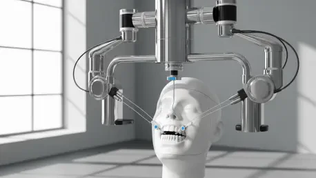

The fundamental challenge of performing high-precision dental surgery lies in the inherent instability of the human mouth, where involuntary movements like breathing, swallowing, and slight head shifts can compromise the accuracy of even the most experienced dental professionals. Unlike the static environments found in industrial manufacturing where robotic arms operate with mathematical certainty on fixed objects, dentistry requires a system capable of adapting to a living, moving target. Traditional robotic solutions have attempted to solve this by using large, external arms paired with complex optical tracking software, but these systems often struggle with latency and the sheer bulk of the equipment. Researchers at the University of Basel have addressed these limitations by developing the Miniature Intraoral Robot, a device roughly the size of a wine cork that mounts directly onto the teeth. By anchoring the robot within the oral cavity itself, the tool moves in perfect synchronization with the patient’s head, effectively neutralizing the problem of movement.
Engineering Innovation: The Distal Drive System
Shrinking a fully functional surgical robot down to a size that can comfortably fit within the human mouth required a significant departure from conventional mechanical engineering. One of the primary obstacles in creating a miniature intraoral device is the management of heat and physical mass, as high-torque motors typically generate enough thermal energy to damage sensitive oral tissues. To circumvent this, the design utilizes a distal drive system where the bulky, heat-generating motors are located outside of the mouth. Power is transmitted to the intraoral component through a series of flexible shafts and cables, allowing the robot to remain lightweight and cool while still delivering the necessary force for dental procedures. This separation of the power source from the surgical site ensures that the workspace remains uncluttered, providing the dentist with a clear view while protecting the patient from the noise and vibrations associated with high-output electrical components.
Mechanical Stability within the Oral Cavity
The mechanical architecture of this system relies on a six-axis configuration that provides the robot with the dexterity needed to navigate the complex topography of different tooth surfaces. By using the patient’s own dental arch as a stable platform, the robot achieves a level of stability that is physically impossible for a hand-held tool or a large external robotic arm. This “inside-out” approach means that any vibration or movement from the patient is automatically shared by the robot, maintaining a constant relative position between the cutting tool and the tooth. This synchronization is critical for tasks like crown preparation, where even a fraction of a millimeter can be the difference between a successful restoration and a damaged nerve. The distal drive cables are engineered to be flexible enough to allow for a wide range of motion while remaining stiff enough to transmit torque with extreme precision, representing a sophisticated balance between mechanical strength and miniature scale.
Digital Workflows: Predictive Dental Restorations
Integrating the Miniature Intraoral Robot into modern practice involves a comprehensive digital workflow that streamlines the entire restorative process from start to finish. In a traditional dental setting, a practitioner must first prepare the tooth by hand before taking a physical or digital impression to send to a laboratory. However, the use of this robotic system allows for a reversal of the typical sequence, where the digital planning occurs before any physical intervention begins. A high-resolution scan of the patient’s mouth is used to create a virtual model, upon which the dentist designs the final crown and the exact path the robot will take to prepare the tooth. Because the robot follows this predetermined map with unwavering accuracy, the permanent crown can be manufactured in advance. This predictive capability means that patients could potentially receive their final prosthetic in a single visit, drastically reducing the time spent in the chair and eliminating the need for temporary restorations.
Material Testing: Validation through Synthetic Models
To validate the structural integrity and functional capability of the robot, engineers subjected the device to rigorous testing using materials that mimic the hardness of human enamel and dentin. The robot performed successful crown preparations on both synthetic resin models and advanced hard hybrid ceramics, demonstrating its ability to maintain consistent cutting forces against resistive surfaces. The procedure follows a two-stage protocol where a larger, high-speed bur is first used for the rapid removal of bulk material, followed by a finer finishing tool to refine the margins and create a smooth surface for the prosthetic. This systematic approach ensures that the final preparation matches the digital design within incredibly tight tolerances. These laboratory trials proved that the miniature actuators and cable-driven system could handle the physical stress of dental drilling without losing calibration, paving the way for the robot to move from a controlled research environment into more complex clinical applications.
Precision DatMotor Encoder Accuracy
Performance data from recent trials indicates that the Miniature Intraoral Robot achieves a level of accuracy that rivals or exceeds the capabilities of manual dental procedures. Analysis of the prepared teeth showed an average positioning error of only 0.18 millimeters, a margin that is remarkably low for a device operating without internal feedback sensors. The robot currently relies on high-resolution motor encoders located at the external drive unit to track its movements, which provides a reliable baseline for precision. Engineers anticipate that the integration of miniature cameras and tactile sensors directly onto the intraoral component will further reduce this error margin, allowing for even more delicate operations. This quantitative consistency is vital for modern dentistry, as it provides a standardized outcome that does not fluctuate based on the fatigue or the varying skill levels of different practitioners. Such reliability is a cornerstone of the shift toward data-driven surgical outcomes.
Clinical Benefits: Conservation of Healthy Tooth Structure
Beyond the technical achievement of precision, the introduction of robotics into the oral cavity supports the growing philosophy of minimally invasive or conservative dentistry. When a dentist prepares a tooth for a crown manually, they often remove additional healthy tissue to compensate for the inherent limitations of hand-held tools and human vision. The robot, however, can follow a surgical path that is optimized to remove only the minimum amount of material necessary to ensure a stable and hygienic fit for the prosthetic. This preservation of the natural tooth structure is essential for maintaining the long-term biological health of the patient, as it keeps more of the enamel and dentin intact. By utilizing digital planning and robotic execution, the dental team can ensure that every micrometer of healthy tissue is protected, leading to better structural outcomes and a reduced risk of post-operative complications or tooth fractures, which are common in over-prepared teeth.
Spatial Constraints: Overcoming Biological Challenges
Transitioning this technology into the messy, liquid-rich environment of the human mouth presents a unique set of clinical challenges that the next generation of robots must solve. While the laboratory tests were conducted in dry or controlled settings, a real-world surgical site is characterized by the constant presence of saliva, blood, and cooling water from the drill. Future iterations of the intraoral robot will need to incorporate advanced fluid management systems to keep the work area clear and protect the internal mechanisms from moisture. Additionally, the limited space in the back of the mouth requires the robot to be even more compact to avoid interfering with the patient’s tongue or cheeks. Researchers have begun designing specialized protective shields and suction attachments that will integrate seamlessly with the robot’s frame. These additions are necessary to ensure that the device remains a practical and safe tool for treating all areas of the mouth, including the notoriously difficult-to-access rear molars.
Future Standards: Real-Time Monitoring and Safety
Looking toward the next phase of development, the primary focus shifted toward the integration of real-time monitoring and autonomous safety protocols. The inclusion of miniature cameras within the robot’s housing allowed for a live feed that the system used to recognize unexpected obstructions or sudden movements. This technological leap enabled the robot to pause its operations instantly if the sensor detected a change in the environment, such as the tongue moving into the path of the bur. The development team also focused on refining the user interface, making it easier for dentists to translate their clinical expertise into robotic instructions without needing specialized programming knowledge. These advancements successfully bridged the gap between a high-tech prototype and a reliable clinical instrument. By prioritizing patient safety and operational efficiency, the project established a new standard for how robotic synchronization and digital planning could be utilized to make dental surgery a faster and more predictable experience.