
For decades, the standard medical protocol for identifying endometriosis has required patients to undergo invasive laparoscopic surgery just to confirm a condition that affects nearly ten percent of the global female population during their reproductive years. This diagnostic bottleneck has historically resulted in an agonizing average wait time of seven to ten years from the onset of symptoms to a formal diagnosis, leaving millions of individuals in a state of clinical limbo. However, as we navigate through the technological landscape of 2026, a paradigm shift is occurring as non-invasive diagnostic tools transition from experimental trials to widespread clinical application. The emergence of high-precision salivary tests and serum-based biomarkers is finally challenging the long-held necessity of surgical intervention for primary diagnosis. These advancements promise to democratize access to early care and fundamentally alter the patient journey by replacing operating rooms with simple collection kits that can be administered in a standard office setting.
Revolutionary Approaches to Molecular Diagnostic Precision
Salivary MicroRNA and High-Throughput Sequencing
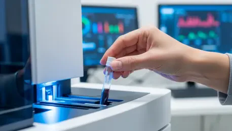
The utilization of microRNA signatures in saliva represents one of the most significant breakthroughs in gynecological diagnostics, offering a sensitivity and specificity that rivals traditional surgical methods. By sequencing small non-coding RNA molecules, researchers have identified specific biological fingerprints that are uniquely associated with the presence of endometriosis, regardless of its clinical stage. In the current clinical environment, products like the Ziwig Endotest have paved the way for a diagnostic protocol that relies on high-throughput sequencing and artificial intelligence to interpret complex molecular data. This method allows for a definitive answer within days rather than years, effectively bypassing the logistical hurdles and physical risks associated with general anesthesia and abdominal incisions. Furthermore, the molecular nature of these tests ensures that even early-stage lesions, which are often missed during visual surgical inspections, can be identified through their distinct epigenetic markers.
Proteomic Profiling and Multi-Marker Serum Panels
Building on the success of transcriptomics, the development of blood-based proteomic assays has introduced another layer of diagnostic certainty for patients experiencing chronic pelvic pain. These tests focus on identifying specific protein concentrations and inflammatory cytokines that circulate in the bloodstream when endometrial-like tissue grows outside the uterus. Unlike previous attempts at blood testing which often yielded low accuracy, the current generation of assays utilizes machine learning to analyze multi-marker panels, providing a comprehensive view of the systemic inflammatory response. This approach is particularly valuable for differentiating endometriosis from other pelvic pathologies such as adenomyosis or irritable bowel syndrome, which frequently present with overlapping symptoms. By providing a clear biochemical signal, these serum tests allow clinicians to initiate targeted hormonal therapies or specialized physical therapy much earlier in the disease progression.
Enhancing Patient Care through Digital Imaging
Artificial Intelligence and Advanced Radiographic Analysis
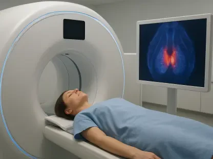
While molecular tests offer a yes-or-no answer regarding the presence of disease, advanced imaging protocols integrated with deep learning algorithms are now providing the anatomical detail necessary for surgical planning. Modern transvaginal ultrasound and specialized magnetic resonance imaging sequences have been refined to detect deep infiltrating endometriosis with a degree of precision that was previously only achievable by expert sub-specialists. Artificial intelligence software now assists radiologists by highlighting subtle distortions in pelvic anatomy and identifying soft signs like reduced organ mobility or obscured tissue planes. This technological assistance reduces the variability between different imaging centers, ensuring that a patient in a rural clinic receives the same diagnostic quality as one in a metropolitan center of excellence. Consequently, imaging has evolved from a tool of exclusion to a vital component of a diagnostic bundle that characterizes the specific phenotype and location of the disease.
Strategic Integration into Modern Healthcare Pathways
The transition toward non-invasive testing necessitated a comprehensive reevaluation of the standard care pathway, shifting the focus from reactive surgery to proactive management. Healthcare systems began to integrate these diagnostic tools into primary care settings, allowing general practitioners to confirm endometriosis during initial consultations rather than referring patients to long specialist waiting lists. This shift effectively eliminated the psychological burden of diagnostic uncertainty and allowed for the immediate implementation of multidisciplinary treatment plans tailored to the specific molecular profile of the patient. The reduction in unnecessary diagnostic surgeries also freed up significant hospital resources, enabling surgical teams to focus exclusively on complex excisions for advanced cases. Moving forward, the medical community emphasized the importance of regular screening for at-risk populations and the continuous updating of AI databases to improve test accuracy across diverse ethnic groups. The end of the diagnostic wait changed the landscape of women’s health forever.