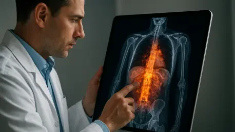

The success of [¹⁸F] AlF-NOTA-Pentixafor in detecting shallow and diffuse gastric tumors represents a pivotal step in refining oncology protocols for indolent lymphomas. Gastric mucosa-associated lymphoid tissue (MALT) lymphoma has long remained one of the most elusive targets in clinical oncology because of its inherently slow growth and low metabolic profile. While traditional diagnostic workflows have leaned heavily on [¹⁸F] FDG PET/CT, this approach frequently fails to distinguish malignant cells from the surrounding healthy tissue. The standard imaging method relies on the high glucose consumption typical of aggressive cancers, yet MALT lymphoma often operates below this metabolic threshold, appearing nearly invisible on a scan. This diagnostic blind spot forces clinicians to rely on invasive biopsies and structural imaging that may not fully capture the extent of the disease. Consequently, staging becomes an exercise in approximation, potentially leading to inadequate treatment plans for patients whose cancer has quietly spread beyond the primary site. The introduction of more sensitive molecular tracers aims to bridge this gap, ensuring that even the most metabolically quiet lesions are brought into sharp focus for medical teams in 2026.
Advancing Diagnostic Precision Through Molecular Targeting
Shifting from Metabolism to Molecular Identity
The fundamental shift in this diagnostic approach lies in moving away from metabolic activity and toward the inherent biological identity of the tumor. The new tracer, known as [¹⁸F] Pentixafor, targets the CXCR4 chemokine receptor, a protein that acts as a critical homing beacon for various immune system cancers. In B-cell malignancies like MALT lymphoma, CXCR4 is frequently overexpressed on the cell surface, playing an active role in the disease’s progression by guiding malignant cells to specific tissue environments and promoting their survival. By targeting this specific receptor, Pentixafor provides a visualization of the cancer’s biological presence rather than just its secondary metabolic effects. This is a significant evolution in nuclear medicine, especially for tumor types where glucose consumption is an unreliable marker for malignancy. Clinicians can now look for the physical signature of the receptor rather than waiting for the cancer to exhibit enough energy consumption to be detected by older, glucose-based tracers.
The biological relevance of CXCR4 extends beyond simple detection, as it is often linked to the aggressiveness and spread of various lymphoid neoplasms. When a tracer specifically binds to this receptor, it provides a more accurate representation of the tumor burden, particularly in cases where the cancer is widespread but not metabolically active. This molecular “identity-based” imaging allows for a level of precision that traditional PET/CT scans simply cannot provide in the context of indolent B-cell lymphomas. By utilizing the [¹⁸F] Pentixafor tracer, oncologists can identify clusters of malignant cells that would otherwise blend in with the normal inflammatory background of the gastric wall. This technique effectively turns a molecular marker into a glowing diagnostic signal, offering a clearer path forward for patients who have previously dealt with ambiguous imaging results. The focus on what the cancer “is” rather than what it “eats” represents a major turning point in how medical professionals approach the management of these complex and deceptive gastric conditions.
Comparative Study Design and Methodology
To evaluate the efficacy of this new molecular tool, researchers at Sichuan Cancer Hospital in Chengdu initiated a prospective, intra-individual study involving twenty patients with confirmed gastric MALT lymphoma. This design was particularly robust because it allowed each patient to serve as their own control, with both [¹⁸F] FDG and [¹⁸F] Pentixafor PET/CT scans performed within a short interval. By comparing the two tracers in the same physiological environment, the research team could eliminate variables such as age, gender, and general health that often complicate large-scale population studies. The study meticulously tracked detection rates and used semiquantitative parameters, such as the maximum standardized uptake value and the tumor-to-blood pool ratio, to provide an objective measurement of the tracers’ performance. This rigorous methodological framework was designed to answer a simple yet vital question: can targeting a specific receptor outperform the current gold standard of metabolic imaging for this specific type of cancer.
The technical execution of the study also involved a deep dive into the statistical significance of the findings, utilizing the McNemar test for paired comparisons to ensure data integrity. The researchers focused not only on the primary gastric site but also on potential secondary sites throughout the entire body. By using high-resolution PET/CT scanners, the team was able to map the exact coordinates of every detected lesion and compare the signal strength between the two different radioactive tracers. This comparative analysis is essential for establishing a new clinical standard, as it provides the hard data necessary to justify a shift in medical practice. The study’s design ensured that every positive signal was cross-referenced with pathological evidence, creating a high-fidelity map of the disease’s presence. Through this exhaustive process, the medical community gained a clearer understanding of how molecular targeting can compensate for the shortcomings of traditional glucose-based imaging in the specialized field of lymphoma diagnostics.
Evaluating Clinical Performance and Practical Application
Analyzing Detection Rates and Signal Strength
The preliminary results of the comparative study revealed a stark contrast in performance, with [¹⁸F] Pentixafor identifying 45% of primary gastric lesions compared to just 20% by the traditional [¹⁸F] FDG method. This more than twofold increase in sensitivity is particularly impressive given the small cohort size and the inherent difficulty of imaging the stomach wall. The tracer provided a much higher signal intensity in the malignant areas, with the median maximum standardized uptake value reflecting a clear distinction between the tumor and the surrounding healthy tissue. This increased clarity is a critical factor for radiologists who must distinguish between early-stage cancer and normal gastric folds or minor inflammation. The higher detection rate suggests that many patients who receive a “clean” scan using traditional methods might actually be harboring undetected disease that is only visible when the CXCR4 receptor is targeted by the new molecular tracer.
Furthermore, the study observed that the signal strength was even more pronounced in extragastric lesions, such as those found in the lymph nodes or lungs. In several cases, [¹⁸F] Pentixafor illuminated secondary disease sites that were completely invisible to the FDG scan, providing a more comprehensive view of the patient’s overall cancer status. This higher uptake in peripheral lesions is vital for accurate staging, as it helps determine whether the disease is localized or systemic. While the statistical significance of the detection rate in this small group was balanced by the researchers’ objective caution, the numerical trend was undeniable. The ability to generate a stronger, more reliable signal in both the primary site and secondary locations positions Pentixafor as a superior tool for monitoring disease progression. This development offers a new level of confidence for oncologists who need to make rapid, life-altering decisions about treatment intensity and the potential for a curative or palliative approach.
Correlation with Structural Changes and Staging
A key aspect of the research involved determining how tracer uptake related to physical changes in the gastric anatomy as seen on standard CT scans. The findings showed a strong statistical correlation between a positive Pentixafor scan and visible gastric wall abnormalities, suggesting that the tracer is highly effective at confirming pathology when structural changes are present. However, the true value of this molecular imaging may lie in its potential to detect disease before it causes significant physical distortion. For patients with MALT lymphoma, early and accurate staging is the cornerstone of effective management. If the disease is truly localized to the stomach, it can often be managed with targeted antibiotics or local radiation. However, if the cancer has spread to distant lymph nodes, the treatment plan must shift toward systemic therapies. A tracer that reliably identifies these hidden spread points ensures that patients receive the correct level of care from the very beginning.
The research also analyzed various clinical predictors, such as age and proliferation indices, finding that none of these factors significantly influenced the tracer’s ability to find the cancer. This suggests that [¹⁸F] Pentixafor’s utility is consistent across a diverse range of patient profiles, making it a versatile tool in the oncological toolkit. By providing a more accurate staging process, the new tracer helps prevent both under-treatment and over-treatment, which is a major concern in the management of indolent cancers. When clinicians can see the full extent of the lymphoma, they can tailor therapies more precisely to the individual’s needs, improving long-term outcomes while minimizing unnecessary side effects. This correlation between molecular signals and clinical staging marks a shift toward a more nuanced understanding of how lymphoma behaves in the human body. The study’s focus on these relationships highlights the importance of integrating advanced molecular imaging with traditional diagnostic techniques to achieve the highest possible accuracy in oncology.
Radiochemistry and Production Feasibility
The practical application of any new medical technology depends heavily on its ease of production and integration into existing workflows, and [¹⁸F] Pentixafor excels in this regard. It is synthesized using an innovative aluminum-fluoride labeling method, which is both efficient and compatible with the standard cyclotron infrastructure found in most modern hospitals. This process avoids the complex and harsh chemical reactions often required for other specialized tracers, making it feasible for routine clinical use. Because it uses the same fluorine-18 isotope as the common FDG tracer, hospitals do not need to invest in expensive new equipment or exotic radioactive materials to begin offering this advanced diagnostic tool. The synthesis process is streamlined and reliable, ensuring that the tracer can be produced on-demand for patients without the logistical hurdles that often plague new radiopharmaceuticals in the early stages of adoption.
The tracer also features a half-life of 110 minutes, which is an ideal window for clinical operations. This duration allows for the tracer to be synthesized, transported to the imaging suite, and administered to the patient, with enough time for the scan to be completed while the signal remains strong. At the same time, the half-life is short enough that the patient’s radiation exposure remains within safe limits and the tracer clears the body relatively quickly. This balance of technical feasibility and patient safety is essential for widespread adoption. Many promising diagnostic agents fail to reach the market because they are too difficult to manufacture or transport, but [¹⁸F] Pentixafor’s reliance on established fluorine-18 chemistry provides a clear pathway to global implementation. As more medical facilities look to upgrade their imaging capabilities in 2026, the logistical simplicity of this tracer makes it an attractive option for improving the care of patients with CXCR4-positive malignancies.
Future Implications for Lymphoma Management
Refining the Paradigm of Cancer Surveillance
The move toward molecularly targeted imaging is fundamentally changing how medical professionals approach the long-term surveillance of indolent lymphomas. In the past, the inability to accurately monitor MALT lymphoma using standard PET scans meant that many patients faced frequent, invasive follow-up procedures like repeat endoscopies and biopsies. The introduction of [¹⁸F] Pentixafor offers a non-invasive alternative that can provide a whole-body view of the disease status with a single scan. This is particularly important for detecting late-term recurrences or transformations that might occur in locations not easily accessible by an endoscope. By shifting the surveillance paradigm from “watch and wait” to “active molecular monitoring,” clinicians can identify changes in the disease earlier than ever before. This proactive approach allows for earlier intervention, which is often the key to maintaining long-term remission in patients with slow-growing but persistent malignancies.
As this technology becomes more common, it will likely be integrated into the standard follow-up protocols for a wide range of B-cell lymphomas. The ability to differentiate between active disease and scar tissue or inflammation is a major advantage of targeting the CXCR4 receptor. In many cases, traditional CT or MRI scans show physical anomalies that are difficult to interpret after a patient has undergone treatment. A molecular tracer that only lights up when it finds specific cancer cells provides the definitive answer needed to clear a patient or restart therapy. This level of diagnostic certainty reduces patient anxiety and helps healthcare systems allocate resources more effectively. The findings from the Sichuan Cancer Hospital study represent just the beginning of this shift, as the medical community continues to explore how these advanced tracers can be used to refine treatment monitoring and improve the overall quality of life for cancer survivors.
Establishing New Standards in Molecular Imaging
The comparative research conducted in Chengdu established a new benchmark for the evaluation of radiopharmaceuticals in the context of rare and indolent cancers. The study successfully demonstrated that [¹⁸F] Pentixafor outperformed the traditional glucose-based tracer in detecting primary and secondary lesions, even within a small and focused patient group. Researchers found that the CXCR4-targeted approach provided a more consistent and clearer signal, particularly in locations where FDG had previously failed. This evidence supported the growing consensus that molecular identity is a more reliable indicator for certain cancers than metabolic activity. By documenting these successes, the study provided the necessary data to encourage larger, multi-center trials that could eventually lead to the formal inclusion of Pentixafor in international oncology guidelines. The move toward “identity-based” diagnostics was validated by the improved detection rates and the tracer’s ability to illuminate the full extent of the disease.
Looking forward, the medical community began to see these results as a call to action for further innovation in the field of nuclear medicine. The focus on specific molecular receptors like CXCR4 opened the door for the development of even more specialized tracers tailored to different types of lymphoma. This targeted approach has the potential to transform oncology from a one-size-fits-all model into a highly personalized discipline. Actionable next steps for the industry include the expansion of manufacturing capabilities for [¹⁸F] Pentixafor and the development of standardized imaging protocols to ensure consistent results across different medical facilities. As larger datasets are compiled, the role of this tracer in the initial staging and ongoing management of gastric MALT lymphoma will likely become the new standard of care. By turning specific biological markers into diagnostic beacons, the healthcare industry moved closer to a future where no cancer remains hidden, regardless of how slow-growing or metabolically quiet it may be.