
When a twelve-year-old girl from Sinaloa, Mexico, began suffering from a persistent cough and severe respiratory distress, neither her family nor her local physicians could have anticipated the extraordinary medical journey that lay ahead. Angela Beltran’s symptoms were initially attributed to common respiratory issues, but as the condition worsened, it became clear that a much more sinister force was at play. Seeking answers that local clinics could not provide, her family traveled to Los Angeles to consult with specialists at Cedars-Sinai Guerin Children’s. There, diagnostic imaging and expertise revealed a three-centimeter mass lodged deep within the bronchus of her left lung. This was not a standard infection or a common pediatric ailment; it was a rare form of cancer that usually appears only in adults. The diagnosis of bronchopulmonary mucoepidermoid carcinoma set the stage for a high-stakes intervention requiring a level of surgical precision that pushed the boundaries of modern pediatric medicine.
Identifying a Rare Pediatric Pathogen
Understanding the Bronchopulmonary Carcinoma
Bronchopulmonary mucoepidermoid carcinoma is a malignancy that originates in the mucus-producing glands of the airways, a condition so exceptionally rare in children that it accounts for fewer than 0.2 cases per every million pediatric tumors. While this type of cancer is occasionally seen in older adult populations, its presence in a pre-adolescent patient presented a unique set of challenges for the oncology team. Dr. Leo Mascarenhas, a specialist in pediatric oncology, recognized that the location of the tumor—lodged within the airway—was particularly precarious because it compromised the patient’s ability to breathe and threatened the long-term viability of the entire lung. Standard protocols for pediatric lung issues often focus on infections or congenital malformations, making the identification of such a specific carcinoma a critical turning point in the case. The rarity necessitated a shift from general pediatric care to a highly specialized approach that integrated the latest oncological research.
Implementing Multidisciplinary Diagnostic Accuracy
To confirm the nature of the growth without causing unnecessary trauma to the young patient’s chest, the medical team employed a multidisciplinary strategy that drew upon both pediatric and adult surgical expertise. Dr. Andrew Brownlee, an adult thoracic surgeon, and Dr. Eugene Kim, the surgeon-in-chief at Guerin Children’s, collaborated to perform a robotic bronchoscopy, which allowed for a minimally invasive biopsy of the mass. This procedure was essential because traditional diagnostic methods might have lacked the precision needed to navigate the narrow pediatric airways while obtaining a sufficient tissue sample for a definitive diagnosis. By utilizing robotic technology at this early stage, the surgeons could visualize the tumor’s exact orientation and its relationship to the surrounding vascular structures. This precise mapping was instrumental in confirming the malignancy and planning a surgical intervention that would prioritize the preservation of healthy tissue while ensuring the complete removal of the cancerous mass.
The Innovation of Robot-Assisted Care
Precision Through Robotic Assistance
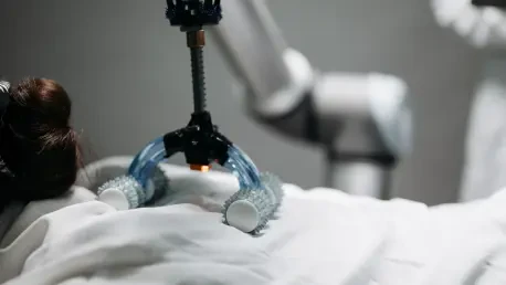
During the complex surgical procedure, the medical team utilized the robotic system to navigate the delicate internal structures of the chest with unparalleled accuracy. The robot’s instruments allowed for the meticulous dissection of the tumor and the subsequent removal of the lower left lobe of the lung, where the malignancy had become deeply embedded. This level of precision was crucial because the bronchus is surrounded by major blood vessels and sensitive nerve pathways that could easily be damaged during a traditional manual procedure. The high-definition 3D visualization provided by the robotic console enabled the surgeons to identify the exact boundaries of the tumor, ensuring that no cancerous cells were left behind while sparing as much healthy lung tissue as possible. This technical capability was particularly vital given the rarity and aggressive potential of the mucoepidermoid carcinoma. The successful isolation and extraction of the mass through minimally invasive ports proved that adult surgical technologies can be safely adapted for younger patients.
Institutional Models: The Future of Specialized Treatment
The integration of robotic technology and collaborative medical models established a new benchmark for treating rare pediatric malignancies with minimal long-term impact. This case demonstrated that when traditional boundaries between adult and pediatric specialties were dissolved, patients benefited from a broader range of technical expertise and advanced resources. Moving forward, healthcare institutions should prioritize the development of similar integrated programs that facilitate the cross-application of surgical innovations across different age groups. Investing in robotic training for pediatric surgeons and fostering partnerships with adult thoracic departments will be essential for improving outcomes in complex cases. The focus must remain on refining these minimally invasive techniques to ensure they are accessible to a wider demographic, particularly for those with rare diseases that fall outside standard clinical protocols. Ultimately, the success of this intervention proved that the fusion of high-definition robotics and specialized care could fundamentally change the trajectory of pediatric oncology.