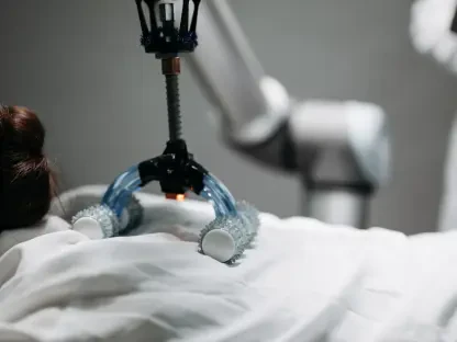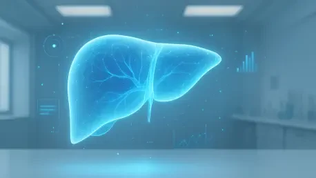

Metabolic dysfunction-associated steatohepatitis represents a severe condition where active fibrosis acts as the primary predictor of liver-related mortality rates. This silent progression of connective tissue buildup, known as fibrosis, has long plagued the medical community because it remains invisible during its most treatable stages. Historically, diagnosing the shift from simple inflammation to permanent scarring required invasive biopsies that only provided a snapshot of a tiny portion of the organ. However, the landscape is shifting toward precision molecular imaging. A newly developed PET tracer, [68Ga]Ga-ATH001, is proving that it is possible to illuminate the “scarring factory” within the body in real-time. By targeting specific receptors on the cells responsible for this damage, clinicians can now distinguish between historical damage and active, ongoing fibrogenesis, offering a window of opportunity for intervention before the damage becomes irreversible and life-threatening. The clinical landscape of chronic disease management is currently witnessing a transformative shift with the emergence of this molecularly targeted imaging, which focuses on visualizing and quantifying fibrosis in conditions like cirrhosis and heart failure.
Clinical Methodology and Patient Trials
Testing the Tracer: Human Subject Trials
The inaugural first-in-human study was spearheaded by a collaborative team of researchers from Uppsala University, the Karolinska Institutet, and several international medical centers. To establish a robust baseline for how the tracer behaves within a living system, the team recruited a small but highly specialized cohort consisting of six participants. This group was split evenly between three healthy volunteers and three individuals already diagnosed with metabolic dysfunction-associated steatohepatitis. This demographic selection was crucial, as it allowed the researchers to compare the tracer’s behavior in a pristine physiological environment versus one burdened by active pathology. By focusing on MASH patients, the study addressed a critical need in hepatology, as these individuals are at the highest risk for progressive liver failure and related complications. The primary goal was to map the biodistribution and safety of the tracer, ensuring that it could be used safely in broader clinical settings. This pilot group provided the essential data required to move from laboratory theories to practical, bedside applications in modern medicine.
Rigorous Experimental Protocols: The Scanning Sequence
To ensure the highest level of data integrity, the experimental protocol followed a strict and detailed timeline. Each participant received a precisely calibrated intravenous bolus of the [68Ga]Ga-ATH001 tracer, utilizing a very low peptide mass of approximately 51 micrograms to minimize any potential biological response while maximizing imaging clarity. The imaging sequence was designed to capture both the rapid initial distribution and the long-term retention of the tracer. It began with an immediate, 35-minute dynamic PET/MRI scan focused on the abdomen to observe how the tracer first interacted with the liver and surrounding tissues. This was followed by a series of whole-body PET scans conducted at one, two, and three hours post-injection. By analyzing 25 different organs and tissues, ranging from the major metabolic hubs like the kidneys to sensitive regions like the red bone marrow, the team created a comprehensive radiation and distribution map. This multi-temporal approach was vital for understanding the tracer’s clearance rates and its ability to bind specifically to target cells over an extended period.
Analyzing Results and Safety Profiles
Distinguishing Health From Disease: Liver Uptake
The results of the biodistribution analysis provided striking evidence of the tracer’s efficacy in distinguishing between healthy and diseased states. In the healthy volunteer group, the tracer cleared rapidly from the bloodstream, and liver uptake returned to near-background levels shortly after the initial injection. Conversely, the MASH patients displayed a significantly different profile, characterized by much higher and more persistent uptake within the liver tissue. Quantitatively, the standardized uptake value in the MASH group averaged 4.0, whereas the healthy group remained much lower at 2.8. This clear statistical separation confirms that the PET tracer successfully identifies the upregulation of PDGFRβ on activated hepatic stellate cells, which are the primary drivers of fibrosis. Effectively, the imaging technology allowed researchers to see the difference between a “quiet” organ and one that is actively being remodeled by disease. This ability to visualize the active “scarring factory” rather than just the final, permanent damage represents a major leap forward in diagnostic accuracy and could eventually replace the need for painful biopsies.
Evaluating Radiation: Clinical Feasibility
For any new radiopharmaceutical to become a standard tool in the clinical arsenal, it must demonstrate a safety profile that allows for repeated use, especially when monitoring chronic conditions over several years. The dosimetry calculations for [68Ga]Ga-ATH001 were highly favorable, yielding a sex-averaged effective dose of roughly 0.028 millisieverts per megabecquerel. When administered at a standard clinical activity level of 150 MBq, the total radiation exposure to the patient is approximately 4.1 mSv, which is significantly lower than the 10 mSv typically delivered during a standard abdominal CT scan. While the kidneys were identified as the dose-limiting organ due to the natural renal excretion and reabsorption of small peptides, the levels remained well within established safety boundaries. Furthermore, none of the participants in the trial reported any adverse effects or discomfort during or after the procedure, even when receiving the highest peptide doses. This level of safety and low radiation burden makes the tracer an ideal candidate for longitudinal studies where patients may need multiple scans to track their progress.
Future Applications and Industry Impact
Expanding Beyond Liver Disease: Multi-Organ Scope
While the initial focus of this research was centered on the liver, the biological target of the tracer, PDGFRβ, is a universal actor in tissue remodeling across the entire human body. This means that the potential applications for [68Ga]Ga-ATH001 extend far beyond hepatology into fields like cardiology and pulmonology. For instance, separate trials are already being planned to investigate whether the tracer can accurately map cardiac fibrosis in patients who have recently suffered a myocardial infarction. In such cases, knowing the extent of active scarring could help cardiologists predict future heart failure risks more accurately. Similarly, the tracer holds immense promise for pulmonary medicine, where it could provide a non-invasive method to monitor the progression of idiopathic pulmonary fibrosis, a condition that is notoriously difficult to track without frequent, high-risk interventions. Even in oncology, the tracer could be used to image the fibrotic stroma that often surrounds tumors, potentially revealing why certain cancers are resistant to chemotherapy and helping doctors to customize treatments more effectively.
Streamlining Drug Development: Clinical Trials
The pharmaceutical industry stands to benefit significantly from the integration of active fibrosis imaging into clinical trial designs. Currently, developing anti-fibrotic medications is an arduous and expensive process because proving a drug’s efficacy often requires serial biopsies taken years apart. This slow feedback loop has hindered the development of new therapies for decades. A validated PET marker for active fibrogenesis, however, would allow researchers to observe a drug’s impact on cellular activity in a matter of weeks. This capability would enable “patient stratification,” a process where doctors can identify which individuals have “active” disease that is likely to respond to a specific treatment versus those with “dormant” scarring that requires a different clinical approach. By providing a real-time readout of biological activity, the tracer could drastically reduce the size and duration of clinical trials, bringing life-saving medications to the market much faster than current methods allow. This shift toward molecular-level monitoring will likely become the new standard for evaluating drug performance starting from 2026.
Redefining the Future of Pathology
A Shift Toward Dynamic Diagnostics: New Paradigms
The successful implementation of [68Ga]Ga-ATH001 marked a pivotal transition in molecular imaging, moving the field from the observation of static, structural changes to the visualization of dynamic, biological processes. Researchers were able to look past the permanent scars of the past and instead focus on the cellular activity occurring in the present. This shift allowed for a much deeper understanding of how chronic diseases actually progress within an individual patient, rather than relying on generalized models of pathology. The study demonstrated that the “scarring factory” could be caught in the act, providing a much more accurate picture of a patient’s current health status than any blood marker or anatomical scan could offer. By offering this new window into human biology, the technology paved the way for a more nuanced approach to diagnosis, where the intensity of the disease process is just as important as the physical damage it has already caused. This evolution in diagnostic philosophy suggested that the future of medicine would rely on seeing the disease as it happens, rather than just treating its long-term consequences.
Early Intervention: Patient Outcomes
The research findings indicated that the ability to detect active fibrogenesis early enough to intervene effectively was finally within reach. By identifying the specific molecular markers of active scarring, clinicians were provided with the tools necessary to move away from reactive treatments and toward proactive, personalized care. The study concluded that catching the scarring process in its early stages could potentially save a vast number of patients from the devastating effects of end-stage organ failure. Looking ahead, the next logical steps involved expanding these trials to larger, more diverse patient populations to further refine the diagnostic thresholds for different organs. Medical professionals were encouraged to consider how these imaging techniques could be integrated into routine screenings for high-risk patients, such as those with chronic metabolic conditions or persistent inflammation. Ultimately, this research did not just present a new way to look at the liver; it established a foundational shift in how chronic organ decay was understood and managed. The transition from invasive procedures to precision scanning represented a significant victory for patient comfort and diagnostic accuracy alike.