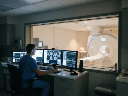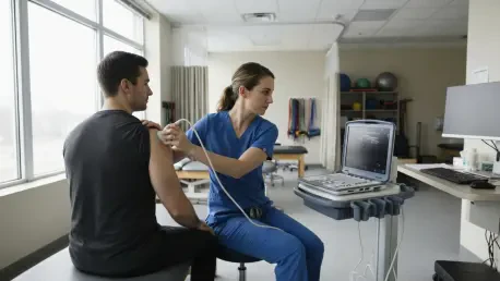

Advanced high-resolution B-mode ultrasound allows for a two-stage screening process that prioritizes muscle trophism over the visually confusing tendon footprint alone. For years, medical professionals have grappled with the structural ambiguity of the rotator cuff, where the supraspinatus and infraspinatus tendons interweave in a complex architecture at the greater tuberosity of the humerus. This anatomical interdigitation often masks the true extent of injury, leading to misdiagnosed tears that are frequently larger than they appear on conventional imaging. When a patient presents with persistent shoulder weakness and limited mobility, the standard diagnostic focus usually lands on the primary tendon insertion points. However, this narrow focus often misses combined lesions that creep into the anterior portion of the infraspinatus. By shifting the perspective toward muscle health—specifically the state of the superior muscle belly—modern imaging reveals hidden patterns of atrophy that act as definitive markers for structural failures. This approach provides a clearer diagnostic roadmap, ensuring that clinicians no longer overlook the critical transitions between distinct muscle units that are vital for shoulder stability and long-term functional recovery.
Clinical Assessment: Physical and Imaging Protocols
Physical Testing: Screening and Preparation
The methodology for detecting these hidden tears begins with a rigorous combination of specialized orthopedic tests and high-resolution B-mode ultrasound. While standard maneuvers such as the Jobe test or Hawkins-Kennedy maneuvers are widely used to assess general impingement and supraspinatus integrity, the Resisted External Rotation Test has emerged as the most critical clinical tool for identifying hidden infraspinatus involvement. To ensure diagnostic accuracy, the clinician must prevent the patient from recruiting synergistic muscles like the posterior deltoid or the teres minor, which are notorious for masking significant muscle weakness. This isolation is achieved by positioning the arm in a neutral rotation while applying consistent resistance, allowing the examiner to feel for subtle drops in power that suggest the superior belly of the infraspinatus is no longer providing its necessary mechanical contribution.
A positive result on these resisted tests, characterized by localized weakness or the inability to maintain force during external rotation, serves as the primary clinical indicator that the superior muscle belly may be compromised. Once this clinical suspicion is established, the diagnostic workflow transitions to a focused ultrasound evaluation that looks beyond the obvious tendon footprint. The goal is to correlate the functional deficit with the physical state of the muscle tissue itself, rather than relying solely on the visual appearance of the tendon fibers. This preparation stage is vital because it filters out general shoulder pain from specific, compartmentalized injuries that require a more nuanced surgical or rehabilitative strategy. By grounding the diagnostic process in reproducible physical examination findings, practitioners can more effectively target the specific anatomical zones that require deeper investigation during the subsequent imaging stages.
Screening Steps: Advanced Ultrasound Techniques
The core of the current diagnostic protocol involves a sophisticated two-stage ultrasound screening process designed to identify muscle trophism and the precise extent of a tear. In the first stage, clinicians perform a sagittal scan of the infraspinous fossa to observe the contour of the superficial fascia. A healthy infraspinatus muscle will typically display a convex or full appearance, indicating robust muscle mass and normal trophism. However, a flattened or concave appearance is an immediate red flag, signaling selective atrophy or hypotrophy in the superior belly. This visual cue is often the first definitive sign that a tear exists, even if the tendon itself appears relatively intact at the greater tuberosity. The ability to visualize this muscle shrinkage in real-time allows for a rapid bedside assessment that can drastically reduce the time between injury and accurate diagnosis.
In the second stage, clinicians utilize axial and longitudinal scans to determine the transverse extent of the lesion, relying on specific bony landmarks to ensure topographic accuracy. One such landmark is the small blunted osseous ridge located on the greater tuberosity, which provides a natural boundary between the superior and intermediate facets. Furthermore, the use of the Van Holsbeeck septum helps differentiate between the supraspinatus and the infraspinatus when the injury is located more proximally. These landmarks are essential because traditional long-axis imaging often fails to capture the three-dimensional complexity of the rotator cuff. By mapping the injury against these stable anatomical features, the ultrasound operator can pinpoint exactly which fibers have been disrupted. This high level of topographic detail is necessary for surgeons who need to know whether to perform a standard repair or a more complex procedure involving the anterior infraspinatus junction.
Muscle Health: Quantifying Reliability and Performance
Strain Elastography: Innovative Diagnostic Metrics
A significant advancement in this diagnostic protocol is the integration of strain elastography to measure the physical stiffness of the muscle tissue. This technology allows clinicians to move beyond qualitative observation by calculating a specific strain ratio. By comparing the deformability of the atrophied superior belly with that of the healthy middle or inferior bellies, practitioners can quantify the degree of tissue degradation. Scientific data indicates that muscle tissue undergoes a physiological loss of compliance and significant stiffening following a chronic tendon tear. Because these changes in tissue elasticity often occur before the structural shrinkage becomes visible on standard B-mode scans, strain elastography acts as a high-sensitivity biomarker for early-stage injury detection.
Furthermore, the calculation of the strain ratio provides an objective, measurable metric that helps standardize the diagnosis of chronic lesions. When the superior belly of the infraspinatus is damaged, it loses its ability to deform under the mechanical pressure applied by the ultrasound transducer, resulting in a lower strain ratio compared to healthy muscle segments. This loss of elasticity reflects underlying fatty infiltration and fibrosis, which are critical indicators of the muscle’s potential for recovery after surgery. By employing this functional mapping technique, clinicians can better predict patient outcomes and tailor physical therapy programs to the specific condition of the muscle tissue. This shift toward quantitative data reduces the subjectivity inherent in visual-only assessments and provides a more robust foundation for clinical decision-making in complex shoulder cases.
Observer Agreement: Statistical Validity and Consistency
The effectiveness of this ultrasound protocol has been reinforced by its high level of reliability and consistency across various patient subjects. In recent clinical evaluations, selective hypotrophy was graded on a standardized scale that accounted for muscle contour, echogenicity, and cross-sectional area. These metrics provided a clear profile of the injury, with mean scores indicating a high degree of consistency in how these hidden tears manifest. Statistical analysis using paired Student’s t-tests further confirmed that the differences in strain ratios between healthy and damaged muscle segments were significant and reproducible. This consistency is vital for integrating the protocol into busy orthopedic practices where multiple clinicians might be involved in a single patient’s care journey.
Most notably, the interobserver agreement for this specific ultrasound methodology reached a perfect Cohen’s kappa coefficient of 1.00. This suggests that the protocol is highly reproducible; provided that clinicians follow the established anatomical landmarks and screening steps, they are virtually guaranteed to arrive at the same diagnostic conclusion. This level of reliability is rare in musculoskeletal imaging, which is often criticized for being overly operator-dependent. Furthermore, comparative studies have shown that findings from this ultrasound method match MRI data with remarkable precision. By providing a low-cost, high-reliability alternative to expensive MRI scans, this protocol democratizes access to advanced shoulder diagnostics, allowing for more frequent monitoring and faster adjustments to treatment plans without compromising the accuracy of the final diagnosis.
Biomechanical Evolution: Insights into Shoulder Stability
Early Warning Systems: The Rotator Cable Role
The research surrounding hidden infraspinatus atrophy has also provided nuanced insights into the role of the rotator cable in shoulder biomechanics. The rotator cable is a thick bundle of fibers that acts like a suspension bridge, distributing mechanical loads across the rotator cuff and protecting the muscle from immediate atrophy in the event of a tear. While this mechanism is highly effective for certain crescent-type supraspinatus tears, it does not appear to provide the same protective shield to the anterior portion of the infraspinatus. The superior muscle belly of the infraspinatus remains highly sensitive to insertional disruptions, which means that even a relatively small tear will trigger observable muscle shrinkage almost immediately after the injury occurs.
This high sensitivity makes the superior muscle belly an ideal early warning system for clinicians who are managing acute shoulder trauma. In cases where a supraspinatus tear might be compensated for by the rotator cable, the state of the infraspinatus provides a clearer picture of the actual mechanical integrity of the cuff. If the superior belly shows signs of flattening or increased echogenicity, it indicates that the injury has progressed beyond the protective range of the rotator cable. Recognizing this early signal allows practitioners to intervene before the muscle undergoes irreversible fatty infiltration. By understanding these biomechanical nuances, clinicians can better interpret imaging results and explain to patients why they might feel significant weakness even if the primary tendon appears to be only partially torn on traditional scans.
Workflow Optimization: Future Directions and Refinement
The ultimate goal of this research was to propose a fundamental shift in how shoulder trauma is managed in clinical settings. Rather than starting the evaluation with the often-confusing tendon footprint, clinicians were encouraged to first screen the infraspinatus muscle for selective atrophy. This screening-led approach ensured that complex, combined tears were accurately identified from the outset. In the recent case series, a finding of concavity in the superior belly served as a definitive red flag that the injury had extended beyond the supraspinatus territory. By prioritizing muscle health as the primary diagnostic indicator, medical teams avoided the common pitfall of misclassifying extensive injuries as simple isolated tears, which frequently led to poor surgical outcomes and prolonged recovery times.
Looking ahead, the transition from operator-dependent strain elastography to absolute quantitative methods like shear-wave elastography was identified as a key step for further refining diagnostic accuracy. Researchers recognized that while current protocols were highly effective, incorporating numeric planimetry for volumetric measurements would provide even more precise data on muscle loss. The findings from 2026 through the subsequent years suggested that standardized ultrasound screening could eventually replace more expensive imaging for routine follow-ups. For the patient, this transition meant more precise surgical planning, more anatomical repairs, and a significantly higher probability of restoring full shoulder function. By integrating these advanced imaging techniques into the daily workflow, the medical community successfully moved toward a more proactive and accurate model of musculoskeletal care.