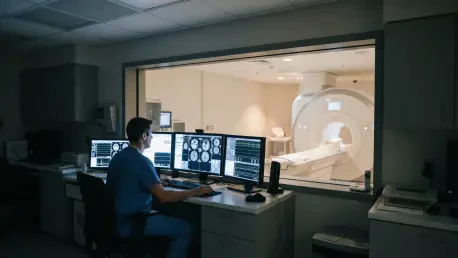

Healthcare facilities currently struggle to balance an exponential surge in patient volume against the limitations of aging imaging infrastructure and restricted staffing levels. This tension is further complicated by the technical nature of Magnetic Resonance Imaging (MRI), where radiologists and technicians must constantly navigate a difficult trade-off between acquisition speed and diagnostic image quality. As demand for rapid diagnostics grows in 2026, many healthcare networks find themselves managing a disparate fleet of scanners that span various manufacturers, field strengths, and chronological ages. This heterogeneity often leads to systemic inconsistencies in software capabilities, which can hamper the efficiency of a centralized diagnostic workflow. Instead of pursuing the traditional and incredibly expensive route of total hardware replacement, forward-thinking institutions are turning toward intelligent software solutions to harmonize their imaging assets and ensure that every patient receives a high-standard scan.
Modernizing Infrastructure: Advanced Software Integration
Bridging the Gap: The Role of Legacy Equipment
To address these multifaceted fleet-wide challenges, imaging centers are increasingly adopting advanced deep-learning applications that function independently of specific hardware providers. These scanner-neutral tools, such as SubtleHD™(MR), allow legacy scanners—some of which have been in service for over a decade—to achieve image quality that rivals the output of much newer, high-field systems. By applying sophisticated denoising and sharpening algorithms to images reconstructed from accelerated scans, this technology effectively decouples software innovation from expensive and slow hardware lifecycles. This fundamental shift enables facilities to modernize their existing assets without the prohibitive capital expenditure typically required for a total hardware overhaul or a magnet replacement. In a period where financial agility is paramount, the ability to refresh a machine’s performance through a software patch represents a revolutionary approach to medical technology management.
This software-centric revolution specifically targets the limitations of older magnets that might otherwise be considered obsolete in a high-throughput clinical environment. By using deep-learning models trained on vast datasets of high-resolution scans, these applications can predict and restore lost detail in images that were previously marred by graininess or low signal-to-noise ratios. Consequently, a machine purchased in the early 2010s can be revitalized to provide the crisp, detailed diagnostic images expected from a 2026-era system. This longevity is crucial for community clinics and satellite offices that may not have the budget for a state-of-the-art 3T installation every few years. The resulting parity across the imaging fleet ensures that clinicians do not have to settle for lower-quality data simply because of a patient’s geographic location or the specific scanner that was available during their appointment, thereby elevating the baseline of care for the entire community.
Implementation Strategies: Scalable and Cost-Effective Solutions
The core benefit of this technology lies in its ability to provide a scalable and repeatable method for fleet modernization without disrupting daily operations. Traditional MRI upgrades often involve replacing massive magnets or entirely reconstructing imaging suites, a process that can take weeks and drain millions from a hospital’s capital budget. In contrast, software-centric optimization allows for a rapid rollout across multiple sites simultaneously, mitigating the noise inherent in faster scan sequences and extending the financial and clinical lifecycle of older equipment. This approach provides a safety net for healthcare administrators, allowing them to allocate funds toward other critical areas like staffing or emergency services while still ensuring their radiology department remains on the cutting edge. By shifting the focus from hardware to software, hospitals can adapt to new clinical protocols much faster than was previously possible in the industry.
Beyond the immediate financial savings, these scalable solutions ensure that radiologists maintain high standards of confidence in their diagnoses, regardless of the machine’s vintage. When noise is reduced and clarity is enhanced via AI, the visual differences between an older 1.5T scanner and a modern 3T system begin to blur, creating a more uniform diagnostic experience. This uniformity is essential for maintaining consistent clinical outcomes and reducing the likelihood of missed findings due to poor image resolution. Furthermore, because the software is often vendor-neutral, it can be applied to a diverse array of machines from different manufacturers, creating a single, unified performance standard across the entire enterprise. This creates a more resilient infrastructure that is less dependent on the specific release cycles of original equipment manufacturers, giving healthcare systems more control over their own technological progression and their long-term operational strategy.
Real-World Applications: Strategic Integration Trends
Case Studies: Performance in Rural Healthcare Settings
The efficacy of these software-driven upgrades was clearly demonstrated by the recent experiences at rural community hospitals like St. John’s Health. For institutions operating with limited capital budgets, the integration of third-party artificial intelligence served as a pivotal shift, allowing a magnet dating back to 2012 to meet and even exceed modern performance standards. By prioritizing software enhancements over hardware replacement, the hospital bridged the performance gap that typically exists between older machines and the latest high-field systems. This strategy allowed the facility to remain competitive in a landscape where patients often travel for better imaging technology. The success at St. John’s illustrated that the age of a magnet no longer dictates the quality of the diagnostic report, provided the underlying data processing is handled by modern algorithms. This realization shifted the internal culture from one of hardware scarcity to one of software-driven abundance.
Clinicians at these facilities often used the analogy of dental hygiene to explain the value proposition to stakeholders, comparing standard manufacturer software to a traditional toothbrush and specialized AI post-processing to an electric version. Both tools are designed to achieve the same fundamental goal, but the latter provides a level of efficiency, depth, and consistency that the standard tool simply cannot match alone. This comparison underscores the prevailing clinical consensus that advanced AI is not merely a replacement for existing tools but a significant enhancement that expands their utility. The transition to an electric-style efficiency in MRI scanning meant that even the most routine studies were performed with a degree of precision that was previously unattainable. This enhancement was particularly valuable for complex cases where subtle details were necessary for an accurate diagnosis, proving that a well-maintained older system could still function as a top-tier diagnostic asset in a modern medical environment.
Synergistic Approaches: Developing Layered AI Pipelines
A significant emerging trend in the imaging sector is the evolution of artificial intelligence from a simple replacement strategy into a more sophisticated and complementary layered framework. Rather than forcing a choice between original equipment manufacturer tools and third-party software, contemporary clinical protocols now favor applying neutral software directly on top of proprietary deep-learning reconstructions. In this synergistic model, the manufacturer’s software handles the initial data acquisition and k-space reconstruction, while the secondary AI layer applies a specialized pass of noise reduction and sharpening. This dual-layered approach creates a cumulative benefit that improves scan speed and image clarity far beyond what either system could realistically achieve in isolation. It represents a move toward an “open” imaging ecosystem where different technologies work together to maximize the potential of the hardware rather than operating in silos.
The practical result of this layered pipeline is a significant reduction in the trade-offs that have historically plagued the radiology field. By combining the strengths of different AI models, technicians can push the boundaries of acquisition speed without fearing a catastrophic drop in image quality. This is especially beneficial for difficult-to-scan patients, such as those with claustrophobia or pediatric patients who struggle to remain still for long durations. The layered AI approach ensures that the first pass of reconstruction is optimized for the specific nuances of the scanner hardware, while the second pass refines the output for the specific diagnostic needs of the radiologist. This collaboration between different software entities creates a more robust and flexible imaging environment, allowing clinics to tailor their workflows to the specific needs of their patient population while maintaining a high baseline of technical excellence across all studies.
Standardizing Outputs: Enhancing Operational Workflow
Cognitive Friction: Reducing Burden Across Multi-Vendor Fleets
One of the most profound benefits of implementing a hardware-agnostic AI layer is its ability to standardize diagnostic output across a diverse imaging network. Radiologists in large systems often review scans from several different sites during a single shift, and varying image quality across different machine brands can lead to significant cognitive friction and diagnostic fatigue. When images from one site are crystal clear and images from another are grainy or filled with artifacts, the doctor must perform a “mental reset” for every new exam. By leveraging an external AI layer, organizations achieve a high level of homogeneity, ensuring that a scan from an older 1.5T system looks substantially similar to one from a brand-new 3T system. This visual consistency removes the need for constant mental recalibration and helps doctors distinguish between genuine pathology and machine-specific artifacts, which speeds up the entire reporting process.
Furthermore, this standardization plays a vital role in reducing the diagnostic errors that can occur when a radiologist is overtired or distracted by poor image quality. When every scan meets a certain aesthetic and technical threshold, the radiologist can focus entirely on the medical findings rather than questioning the integrity of the data. This streamlining of the visual experience is a key factor in improving daily workflow and overall job satisfaction for highly specialized medical staff. In an era where burnout is a major concern in the healthcare industry, any tool that reduces the mental load of interpreting difficult images is seen as a major operational victory. By creating a unified “look and feel” for every scan in the enterprise, the software-driven approach fosters a more cohesive and efficient diagnostic environment, ultimately leading to faster turnaround times for patients and more reliable results for referring physicians.
Quantifiable Metrics: Maximizing Total System Efficiency
The integration of this AI strategy has produced measurable benefits across five specific pillars of value: acquisition efficiency, workflow acceleration, scheduling capacity, financial performance, and patient experience. By combining manufacturer AI with neutral software layers, facilities have yielded average scan time reductions of approximately 15% per patient, which translates to a saving of several minutes per exam. For high-volume protocols—such as brain scans for stroke assessment or orthopedic studies of the knee—scan times have been reduced by nearly 50% in some clinical environments. These efficiencies allow facilities to add one or two extra patient slots to their daily schedule, directly addressing the backlog of appointments and boosting the bottom line. With net revenues for MRI studies remaining a significant part of hospital income, this increased throughput is a powerful argument for the adoption of software-driven optimization.
Beyond the financial metrics, the improvement in the patient experience is equally quantifiable and impactful. Faster scan times mean that patients spend significantly less time inside the narrow, noisy bore of the magnet, which reduces the likelihood of patient movement and the subsequent need for repeat scans. This reduction in motion artifacts not only improves the data quality but also prevents the frustration of redos that can disrupt an entire day’s schedule. When patients are more comfortable, they are more likely to comply with instructions, leading to a smoother workflow for the technicians on the floor. The combination of financial gains and improved patient satisfaction creates a compelling case for software-driven AI as a foundational component of modern radiology management. By maximizing the utility of existing assets, healthcare systems are finding they can do more with less, turning their aging fleet into a high-performance engine for diagnostic excellence.
Strategic Evolution: The Future of Enterprise AI
Fleet Management: Transitioning to Foundational Systems
The transition of enterprise artificial intelligence from a temporary patch for aging hardware into a foundational fleet management strategy was completed by mid-2026. Clinical leaders recognized that a unified, vendor-neutral software layer was no longer a luxury but an essential component for maximizing scanner utilization across the board. This shift allowed healthcare networks to maintain cross-facility consistency without being tethered to the slow and expensive cycles of hardware replacement. By adopting this unified approach, organizations established a predictable path for technological growth that was independent of which manufacturer they chose for their next magnet purchase. This flexibility empowered administrators to make purchasing decisions based on clinical need rather than software compatibility, fostering a more competitive and innovative marketplace for imaging equipment. The strategy ensured that the entire fleet remained current with the latest diagnostic standards.
As medical imaging continued its digital transformation, the role of the vendor-neutral layer became the primary driver of operational efficiency. Institutions that embraced this model found they could improve an existing system beyond its original specifications, ensuring they remained competitive in an increasingly demanding clinical environment. This ability to “future-proof” existing magnets meant that the traditional seven-to-ten-year replacement cycle was effectively extended, allowing for a better return on investment for every piece of hardware. The long-term implications of this shift were profound, as it allowed for a more sustainable approach to technology management that prioritized software agility over hardware bulk. By the end of this transition, the most successful imaging departments were those that viewed their scanners as versatile platforms for continuous software improvement rather than static pieces of medical equipment.
Operational Sustainability: Securing Financial and Clinical Success
The ultimate finding from the broad implementation of these technologies was that software-driven AI became a financial and clinical necessity for high-performing departments. By adopting a layered AI approach, institutions delivered superior patient care while significantly improving their operational throughput. This strategy ensured that even the oldest machines in a fleet contributed to a high-volume, high-quality diagnostic environment, which was essential for meeting the rising patient demands of 2026. The financial sustainability of radiology departments was bolstered by the ability to squeeze more performance out of existing assets, providing a viable path toward long-term healthcare delivery. Leaders who prioritized these upgrades saw a marked improvement in both staff retention and patient outcomes, as the stress of managing outdated equipment was replaced by the confidence of working with modern, AI-enhanced tools.
As the industry moved forward, the actionable takeaway for stakeholders was the importance of decoupling software innovation from hardware procurement. Organizations that invested in a robust, scanner-neutral AI layer were better positioned to react to new medical challenges and shifts in reimbursement models. This flexibility proved to be a major competitive advantage, allowing facilities to pivot their clinical focuses without waiting for a multi-million dollar magnet upgrade. The success of this model has paved the way for future developments in automated diagnostics and remote imaging support, further solidifying the role of software as the brain of the modern radiology suite. Looking ahead, the focus will likely remain on refining these digital tools to achieve even greater levels of automation and precision, ensuring that diagnostic imaging remains a reliable and efficient pillar of the global healthcare system for years to come.