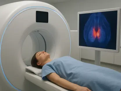

Urologists are increasingly looking toward theranostics to resolve the clinical dilemma of equivocal MRI findings that currently lead to a high volume of redundant tissue samplings. This shift reflects a broader commitment to precision medicine, where the objective is to replace invasive, generalized protocols with highly specific diagnostic pathways. For decades, the standard of care for suspected prostate cancer has followed a predictable and often painful trajectory: an elevated prostate-specific antigen (PSA) test leads to a multi-parametric magnetic resonance imaging (mpMRI) scan, which then frequently triggers a transrectal or transperineal biopsy. While mpMRI has improved detection rates, it often produces results that sit in a gray area, neither confirming nor ruling out malignancy. In such cases, the default clinical response is almost always a biopsy, even though a significant majority of these procedures do not reveal aggressive disease. The arrival of advanced imaging agents like Illuccix and Gozellix represents a pivotal change in this narrative, offering a functional look at cellular activity that structural imaging simply cannot provide. By moving these scans earlier into the diagnostic sequence, clinicians hope to create a filter that captures only the most relevant cases for intervention, effectively reducing the physical and psychological burden on patients who would otherwise face unnecessary surgical sampling.
Enhancing Diagnostic Accuracy Through Advanced Imaging
Redefining the Role: PSMA-PET Scans
The current diagnostic framework typically utilizes Prostate-Specific Membrane Antigen (PSMA) imaging only after a cancer diagnosis is confirmed, primarily to detect metastasis or recurrence. However, the paradigm is shifting to move this powerful technology “upstream” as a primary diagnostic tool. By integrating PSMA-PET scans earlier in the process, physicians gain access to biological data that structural imaging like MRI simply lacks. While an MRI provides a detailed map of the prostate’s anatomy, it cannot always distinguish between inflammation and high-grade cancer. PSMA-PET imaging works by using a radioactive tracer that binds specifically to the PSMA protein, which is overexpressed on the surface of most prostate cancer cells. When these tracers, such as Gallium-68 or Fluorine-18, illuminate a specific area, they provide functional evidence of disease. This level of biological insight allows for a more nuanced interpretation of suspicious lesions, particularly those that appear ambiguous on standard scans, thus providing a clearer roadmap for subsequent clinical decisions.
The methodology behind this diagnostic evolution is currently being solidified through large-scale clinical research, most notably the BiPASS study. This prospective, open-label trial is enrolling 350 patients across multiple high-volume centers in the United States and Australia to evaluate the efficacy of PSMA-PET in the pre-biopsy setting. The study specifically targets men with a “suspicious” MRI finding, categorized as PI-RADS 3, 4, or 5, to see if the addition of a PET scan can more accurately predict which patients harbor clinically significant prostate cancer. By comparing the imaging results directly with the eventually collected pathology, researchers are building a robust evidence base to justify a change in the global standard of care. This approach is not merely about finding more cancer, but about finding the right cancer—the aggressive, fast-growing cells that require immediate treatment—while safely monitoring low-risk cases that might never cause harm to the patient during their lifetime.
Addressing the High Rate: Unnecessary Procedures
The urgent need for a more refined diagnostic triage is underscored by the staggering number of negative biopsies performed every year. Current data suggests that of the millions of prostate biopsies conducted annually, approximately 75% show no sign of significant malignancy. This means that a vast majority of men are undergoing a procedure that is not only invasive but also carries a significant risk profile. Complications such as severe infection, sepsis, rectal bleeding, and urinary retention are not uncommon, and the psychological impact of a “false alarm” can lead to long-term anxiety. By implementing a high-specificity imaging filter like PSMA-PET before the needle ever touches the patient, the medical community aims to drastically reduce these “redundant” procedures. This logic suggests that if the imaging shows no significant PSMA uptake, the likelihood of finding high-grade cancer is so low that a biopsy can be safely deferred in favor of active surveillance.
Beyond the clinical risks, the current biopsy-heavy model faces significant challenges regarding patient compliance and resource management. Statistics indicate that roughly 25% of men who are recommended for a biopsy actually decline the procedure, often due to fear of the associated pain or potential side effects like erectile dysfunction or incontinence. This avoidance can lead to dangerous delays in diagnosis for those who do actually have aggressive disease. Furthermore, the financial burden of these unnecessary surgeries on the healthcare system is substantial. By shifting resources toward advanced imaging—a non-invasive, outpatient procedure—healthcare providers can ensure that surgical interventions are reserved for those who will benefit most. This transition toward a precision-based “triage” system is expected to improve the patient experience by offering a less intimidating path to a definitive diagnosis while simultaneously optimizing the allocation of hospital resources.
Clinical Foundations and Regulatory Milestones
Building on Success: Preliminary Studies
The momentum behind the “imaging-first” approach is not based on theory alone but is supported by data from foundational trials known as PRIMARY and PRIMARY2. These studies acted as the proof-of-concept for the current clinical shift, demonstrating that the combination of mpMRI and PSMA-PET significantly outperformed MRI alone in identifying high-grade prostate cancer. Specifically, the results indicated that the sensitivity and negative predictive value of this dual-modality approach were high enough to potentially eliminate up to half of all unnecessary biopsies. These findings were particularly impactful for patients with PI-RADS 3 or 4 scores on their MRI—the “gray area” where clinical uncertainty is highest. By providing a secondary layer of biological verification, the PET scan allowed urologists to confirm the presence of significant disease with a degree of confidence that was previously unattainable without surgical sampling.
As the medical community moves through 2026, the focus has shifted toward validating these results in the Phase 3 BiPASS trial to satisfy the rigorous requirements of regulatory bodies. Urologists and oncologists are increasingly vocal about the need for this technology to be integrated into official clinical guidelines. The goal is to move beyond “expert opinion” and toward a data-driven standard of care where PSMA-PET is recognized as an essential component of the initial workup. This transition is essential for changing the mindset of practitioners who have relied on PSA and MRI as the primary gatekeepers for decades. By demonstrating that PSMA-PET can reliably identify patients who can safely avoid the biopsy suite, these trials are paving the way for a more sophisticated, less reactive form of oncology that prioritizes both diagnostic accuracy and patient quality of life.
Paving the Way: Global Access and Reimbursement
A significant milestone in the journey toward widespread adoption was the recent alignment between clinical researchers and the United States Food and Drug Administration (FDA) regarding the regulatory pathway. This agreement on a New Drug Application (NDA) pathway ensures that the data collected in current trials will be directly applicable to the approval of PSMA-PET tracers for pre-biopsy use. This regulatory clarity is a vital strategic step, as it provides a clear timeline for when this technology can move from specialized research centers into community urology practices. Without this formal approval, the use of PSMA imaging for primary diagnosis would remain “off-label,” limiting its availability to the general public and hindering the development of standardized protocols that are necessary for high-quality care across different healthcare settings.
Equally important to regulatory approval is the establishment of a robust reimbursement framework. In the current healthcare landscape, the cost of advanced PET imaging can be a barrier for many patients if it is not covered by insurance or national health systems. The ongoing efforts to secure “new product” reimbursement status aim to make these scans a routine, affordable part of the diagnostic journey. By proving the cost-effectiveness of this approach—specifically how the cost of a PET scan is offset by the savings from avoided biopsies and treated complications—advocates are making a strong case to payers. The vision is to ensure that precision diagnostics are not an elite luxury but a standard tool available to all men, regardless of their socioeconomic status or geographic location. This economic integration is the final hurdle in making the “imaging-first” model a global reality that benefits the entire population.
The Future of Prostate Cancer Management
Integrating Diagnosis: Targeted Therapy
The evolution of pre-biopsy imaging is deeply intertwined with the rise of theranostics, a field that seeks to unify the diagnostic and therapeutic stages of cancer care. The same PSMA proteins that are targeted by imaging agents like Gozellix are also the targets for advanced radiopharmaceutical therapies, such as Lutetium-177 PSMA-617. This means that the initial diagnostic scan does more than just confirm the presence of cancer; it also “maps” the targets for future treatment. If a scan reveals significant disease, the patient can transition seamlessly into a targeted therapy protocol that uses the same biological pathway to deliver radiation directly to the cancer cells while sparing healthy tissue. This end-to-end approach represents the pinnacle of personalized medicine, where the diagnostic process is not a separate hurdle to clear, but the first step in a cohesive, biology-driven treatment plan.
This integration is expected to fundamentally change how prostate cancer is managed over the next few years. As diagnostic scans become more sensitive, the medical community will be able to detect and treat smaller clusters of cancer cells before they have the chance to spread or cause symptoms. This “see-and-treat” model reduces the need for the aggressive, whole-organ surgeries or broad-spectrum radiation treatments that have traditionally defined prostate cancer care. Instead, clinicians can move toward a model of focal therapy, where only the identified lesions are treated, preserving the surrounding healthy tissue and maintaining the patient’s functional health. The synergy between high-precision imaging and targeted therapy is creating a future where the management of prostate cancer is less about invasive intervention and more about the intelligent application of molecular science.
Evolving Standards: The Path Forward
The clinical community reached a consensus that the successful validation of PSMA-PET imaging in the pre-biopsy setting marked a definitive shift in urological oncology. Moving forward, the focus was placed on the practical implementation of these findings within the broader healthcare infrastructure. This transition required not only the availability of tracers but also a significant investment in clinician training and the expansion of PET-CT imaging facilities. Radiologists and urologists collaborated more closely than ever before, developing unified reporting systems to ensure that the functional data from PET scans was interpreted with the highest degree of consistency. This collaborative effort ensured that the transition from a biopsy-first to an imaging-first model was supported by a workforce capable of navigating the complexities of molecular diagnostics.
Furthermore, the integration of advanced imaging necessitated a re-evaluation of patient counseling and shared decision-making. Physicians were tasked with explaining the nuances of PSMA uptake to patients, shifting the conversation from a binary “cancer or no cancer” to a more detailed discussion about the biological activity of the disease. This empowered patients to take a more active role in their care, particularly when deciding between active surveillance and intervention. The move toward a non-invasive diagnostic triage proved to be a major victory for patient safety and healthcare efficiency. As these protocols became the global standard, the incidence of biopsy-related complications plummeted, and the detection of truly significant, life-threatening cancers improved, solidifying the role of precision imaging as the cornerstone of modern prostate cancer management.