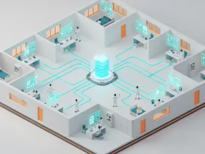

The medical community is currently witnessing a transformative shift in molecular imaging through the development of a portable positron emission tomography system that eliminates the need for massive, lead-shielded facilities. Traditionally, obtaining high-resolution metabolic data required transporting critically ill patients through hospital corridors to a centralized imaging suite, a process fraught with logistical challenges and clinical risks. However, recent breakthroughs by researchers at Washington University in St. Louis have introduced a mobile alternative that brings the diagnostic power of PET technology directly to the patient’s bedside. This innovation is not merely about shrinking the hardware; it represents a fundamental change in how clinicians approach diagnostic procedures in intensive care units and operating rooms. By prioritizing accessibility and real-time data, this system addresses a significant gap in modern medicine, allowing for continuous monitoring and immediate intervention in ways that were once considered physically impossible within the confines of a standard hospital ward or surgical suite.
Engineering Versatility: The Robotic Arm Architecture
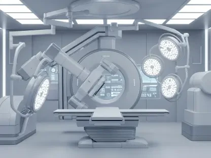
The engineering philosophy behind this mobile system centers on a radical departure from the traditional circular gantry that has defined medical imaging for decades. Rather than forcing a patient into a fixed tunnel, this new design utilizes a highly sophisticated robotic arm that supports modular detector panels, granting the medical team unparalleled freedom of movement during a scan. This “arbitrary positioning” capability means that the detectors can be angled and rotated into specialized configurations that accommodate the myriad of tubes, monitors, and life-support equipment often surrounding a patient in a critical care setting. Such a flexible architecture ensures that the imaging process does not interfere with the essential physical space required for patient care, effectively turning any room into a temporary high-tech diagnostic suite. The robotic arm functions with high precision, moving panels in concert to capture the necessary photons from radioactive tracers while remaining unobtrusive to the clinicians who are actively managing the patient’s condition.
In the high-pressure environment of an interventional radiology suite, physical space is a premium commodity that dictates the feasibility of certain medical procedures. Standard PET scanners are notoriously bulky and immobile, creating a physical barrier that often forces doctors to choose between real-time metabolic imaging and ease of physical access to the surgical site. The introduction of a benchtop, robotic-arm-based system solves this dilemma by allowing the detectors to slide into the procedural field for quick data acquisition and then retract just as easily when the physician needs to resume manual tasks. This fluid movement allows for a symbiotic relationship between imaging and intervention, where the technology serves as a guidance tool rather than a logistical obstacle. Consequently, surgeons can now perform complex maneuvers or biopsies without fearing that they might bump into heavy machinery, while still benefiting from the metabolic context that only a positron emission tomography system can provide during a live operation.
Accelerating Insights: Incremental Reconstruction Algorithms
Processing the massive volume of raw data generated by a PET scan usually requires a significant lag time before a physician can view a final, diagnostic-quality image. To overcome this hurdle, the research team developed a proprietary incremental reconstruction algorithm that functions as an interactive workflow, building the image bit by bit in real time. Unlike traditional methods that process data in a single batch after the scan is finished, this software analyzes information as it is collected, allowing the visual output to refine and sharpen while the detectors are still moving. This immediate feedback loop provides clinicians with a growing window into the patient’s internal metabolic state, enabling them to make decisions faster than ever before. If a specific area of interest is identified early in the scan, the doctor can focus the procedure on that region immediately, rather than waiting for post-scan processing. This software-driven approach effectively turns molecular imaging into a live feed.
Clinical evaluations of this real-time imaging capability have revealed that high-quality features often become diagnostic-grade after only a minimal number of detector movements. This finding has profound implications for patient safety and hospital efficiency, as it allows medical professionals to terminate a scan the moment the necessary diagnostic information is obtained. By reducing the overall duration of the imaging session, the system significantly limits the patient’s exposure to radiation and the time they must remain stationary, which is particularly beneficial for pediatric or highly unstable patients. Furthermore, shortening the scan time increases the throughput of the imaging department, allowing more procedures to be performed in a single day without sacrificing the quality of care. This efficiency ensures that the bedside system is not just a convenience, but a high-performance tool that optimizes the use of hospital resources and radioactive tracers, making the entire diagnostic cycle more sustainable.
Enhancing Precision: Interventional Oncology Procedures
While conventional imaging modalities like X-rays and computed tomography are indispensable for visualizing detailed anatomy, they often fail to capture the underlying biological activity of a tumor. The bedside PET system bridges this gap by highlighting metabolic “hot spots,” which are areas where cells are consuming energy at an accelerated rate, typically indicating aggressive cancer. In the context of interventional radiology, having this metabolic map available at the bedside allows for far more precise needle placement during biopsies. Instead of targeting a lesion based solely on its shape or size, doctors can now aim for the most biologically active portion of the mass, which significantly increases the likelihood of a successful and informative tissue sample. This precision reduces the need for repeat procedures and provides oncologists with a much clearer picture of the tumor’s character from the very beginning of the diagnostic journey, leading to more personalized and effective treatment plans for patients.
The real-time nature of this technology provides a critical advantage during specialized tumor treatments, such as thermal ablations, where heat is used to destroy malignant cells. Traditionally, doctors had to wait hours or even days to perform a follow-up PET scan to confirm that a treatment successfully neutralized the target area. With a portable system, however, the medical team can verify the results of the ablation immediately after the procedure is finished, while the patient is still on the table. If the imaging reveals residual metabolic activity, the interventionalist can apply additional treatment right then and there, rather than scheduling a secondary surgery weeks later. This immediate confirmation loop not only improves the overall success rate of cancer treatments but also reduces the physical and emotional burden on patients who might otherwise face a prolonged period of uncertainty. This capability marks a shift from reactive to proactive surgical imaging, ensuring the job is done right the first time.
Democratizing Access: Mobile Infrastructure Benefits
Traditional PET technology is synonymous with massive financial investments and complex infrastructure requirements that often limit its availability to major urban medical centers. Installing a standard scanner usually involves reinforcing hospital floors to support several tons of equipment, installing specialized cooling systems, and dedicating a large amount of square footage to lead-shielded rooms. In contrast, the portable benchtop unit operates on a much smaller scale, dramatically reducing the overhead costs associated with molecular imaging. Because it does not require a permanent, high-shielded installation, this mobile unit can be shared among different departments or even moved between satellite clinics, bringing high-end diagnostics to a wider range of patients. This flexibility effectively democratizes access to precision medicine, allowing smaller healthcare facilities to offer metabolic imaging services without the multi-million dollar price tag typically required for a dedicated suite, thereby improving the standard of care.
A common concern when downsizing medical equipment is whether the reduction in physical size leads to a corresponding decrease in diagnostic performance or image resolution. Rigorous comparative studies have addressed this issue by demonstrating that the images produced by the portable prototype are as sharp and reliable as those generated by full-sized, stationary PET scanners. The sophisticated sensors and optimized algorithms used in the compact unit ensure that the sensitivity and spatial resolution remain high enough to detect small or subtle metabolic changes in the tissue. This performance parity means that clinicians do not have to compromise on data quality when choosing the portable option for bedside imaging. By maintaining these high standards, the researchers have ensured that the mobile system is a legitimate clinical tool rather than a lower-tier alternative. This reliability is essential for gaining the trust of the medical community and facilitating the widespread adoption of the technology across various clinical specialties.
The Implementation: Navigating Clinical Standardization
As the development project transitions from the laboratory prototype phase toward full clinical integration, the research team is placing a significant emphasis on human ergonomics and operational simplicity. The goal is to create a user interface that is intuitive for clinicians who are not necessarily experts in nuclear medicine, such as intensive care nurses or general surgeons. By simplifying the interaction between the operator and the machine, the technology can be more seamlessly incorporated into the fast-paced environment of the hospital. Current efforts involve refining the software to automate many of the calibration and positioning tasks, allowing the medical staff to focus more on patient care rather than the mechanics of the imaging process. This focus on usability ensures that the system will be practical for everyday use once it enters the final stages of clinical validation, moving beyond its role as a research tool to become a staple of modern medical diagnostics that can be utilized effectively by a variety of departments.
The research team successfully established a clear timeline for the first human imaging studies, which were planned to begin by 2027. They prioritized the development of standardized protocols that allowed the portable PET system to fit into existing clinical workflows without disrupting established safety procedures. This groundwork provided a blueprint for how future medical centers could integrate molecular imaging into their routine bedside care, potentially transforming oncology and critical care medicine. By demonstrating that high-resolution metabolic data was achievable in a mobile format, the developers opened the door for new research into real-time drug monitoring and the treatment of acute neurological conditions. Ultimately, the project moved the medical community toward a more agile diagnostic model where the equipment adapted to the patient’s needs, rather than the patient adapting to the limitations of the facility. These advancements suggested that the next phase of healthcare would be defined by the ubiquity of high-precision tools in any clinical environment.