
The success of a modern neurovascular intervention often depends on a margin of error thinner than a human hair, making the absolute stability of the patient the most critical variable in the operating room. In high-pressure clinical settings such as stroke treatment or complex oncology procedures, every millimeter of accuracy and every second of time saved translates directly into improved patient outcomes and significantly reduced mortality rates. The conventional model of healthcare requires the patient to be physically repositioned to meet the imaging requirements of fixed hardware, but this approach introduces significant risks to procedural integrity and safety. By transitioning to a paradigm where the imaging system moves fluidly around a stationary patient, clinical teams can maintain an uninterrupted focus on the operative site while the technology adapts to the specific needs of the individual. This shift represents more than just a mechanical upgrade; it is a fundamental redesign of the clinical workflow centered on the principles of stability, precision, and streamlined care.
The Critical Risks of Patient Displacement
Precision: Navigational and Clinical Hazards
Maintaining a static patient position is the cornerstone of modern image-guided navigation, particularly when utilizing sophisticated 3D roadmapping software. In neurovascular interventions, clinicians often work with vessels that are only a few millimeters in diameter, requiring sub-millimeter precision for the safe deployment of catheters and micro-stents. When a procedure necessitates moving the patient to achieve a better imaging angle, the delicate alignment between the pre-acquired roadmap and the live fluoroscopic feed is immediately compromised. This misalignment forces the medical team to re-acquire images, which not only consumes precious time during a critical procedure but also increases the patient’s exposure to ionizing radiation and contrast media. Furthermore, the act of moving a patient who already has instruments inside their vasculature risks mechanical complications such as vessel perforation or catheter dislodgment, which can lead to life-threatening internal hemorrhaging within the suite.
Safety: Physiological and Environmental Risks
Many patients undergoing interventional procedures are in a hemodynamically unstable or fragile physiological state, often requiring continuous monitoring and heavy sedation. Repositioning these individuals increases the risk of serious physical complications such as skin shearing, nerve compression, or the accidental dislodgment of life-sustaining tubes and lines. For patients under general anesthesia, any sudden movement can interfere with the airway management or the stability of the breathing circuit, creating a secondary emergency in the middle of a primary vascular intervention. Additionally, the labor-intensive process of moving a patient often requires multiple staff members to crowd around the procedural table, which can lead to accidental contamination of the sterile field. Maintaining the patient in a single, fixed position minimizes the need for physical contact and staff movement, thereby significantly reducing the incidence of healthcare-associated infections and protecting the patient’s stability.
Pioneering the Patient-Centric Environment
Integration: Flexible Imaging and Room Design
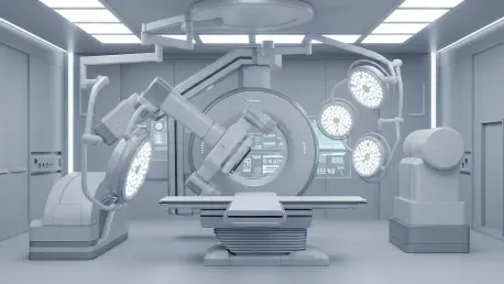
Advanced robotic C-arm systems represent the pinnacle of modern imaging technology, offering unparalleled maneuverability that allows for full anatomical coverage without moving the patient. These systems are designed with multiple axes of motion, enabling the gantry to rotate, tilt, and pivot around the procedural table to capture high-definition images from any necessary angle. This flexibility is particularly vital in multi-disciplinary suites where clinicians may need to switch between imaging the cranial vault and the peripheral arteries in a single session. The ability of the technology to seamlessly transition through these complex movements ensures that the medical team can maintain a constant visual on the target area. Furthermore, the synchronization between the imaging hardware and the table’s software allows for automated positioning, where the system remembers specific angles, reducing the cognitive load on the operator and ensuring that the most effective views are always just a touch away.
Efficiency: The Single-Platform Pathway and Workflow
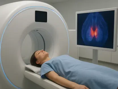
The implementation of a single-platform pathway is a revolutionary step toward optimizing the continuum of care for patients requiring urgent interventional therapy. In this model, the patient is placed on a specialized procedural table upon arrival and remains on that same surface throughout the entire imaging, diagnostic, and treatment phases. This eliminates the need for multiple patient transfers between transport trolleys and various imaging tables, which are frequent points of injury and equipment failure. Reducing these transfers is especially critical for patients with suspected spinal injuries or those undergoing active thrombolytic therapy, where any unnecessary movement could exacerbate their condition. The single-platform approach also streamlines the workflow for the hospital staff, as it reduces the time spent on logistics and preparation. By keeping the patient stationary and moving the technology to them, the medical facility can significantly decrease the critical door-to-needle time in high-stress cases.
Optimization of Safety and Efficiency
Protection: Maximizing Workflow and Radiation Safety
One of the most critical advantages of moving the imaging system instead of the patient is the significant reduction in radiation exposure for both the clinical team and the patient. When a system can be precisely and quickly positioned at the optimal angle, the need for scout images or trial fluoroscopy runs is greatly minimized. This efficiency directly impacts the total screening time, adhering to the ALARA principle which is the gold standard in radiation safety. Advanced robotic systems also allow for better integration of radiation shielding, as the shields can be moved in tandem with the gantry to provide continuous protection without interfering with the procedural access. For medical professionals who spend several hours a day in the interventional suite, this reduction in cumulative scatter radiation is a vital safeguard against long-term occupational health risks. By prioritizing a system that moves with precision, the facility ensures that the benefits of imaging are achieved with the lowest possible dose.
Strategic Advancements: The Path Forward
The transition toward imaging systems that prioritized the stability of the patient over the convenience of hardware marked a significant milestone in clinical safety. Medical facilities that adopted these robotic and mobile gantry solutions observed a measurable decline in procedural complications and an increase in the accuracy of delicate vascular interventions. It was found that maintaining a stationary patient environment allowed for a more disciplined application of sterile protocols and a reduction in the time required for complex image acquisition. To build on these advancements, healthcare administrators were encouraged to evaluate their current suite configurations and prioritize the acquisition of adaptable hardware. Future investments were directed toward systems that offered seamless integration with digital navigation tools, ensuring that the technology remained a dynamic partner in the operating room. By focusing on the mobility of the system, hospitals successfully enhanced the safety, efficiency, and overall quality of the care they provided.