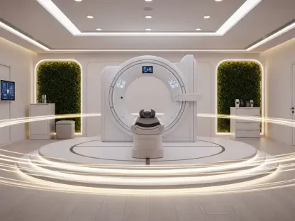

The rapid evolution of Optical Coherence Tomography has transformed it from a niche ophthalmological tool into a versatile powerhouse capable of revealing the microscopic secrets of human tissue across various medical specialties. While clinicians have long relied on this technology to map retinal architecture with unparalleled precision, the current landscape of medical imaging is pushing these boundaries into the realms of oncology, dermatology, and even complex dental procedures. The core value of this modality lies in its ability to capture raw signals that contain a wealth of hidden information regarding the biophysical state of tissue, such as light absorption rates and subtle phase alterations. However, the vast majority of this diagnostic potential remains locked away because the raw data is notoriously difficult to interpret without specialized computational tools. Historically, the transition from simple structural visualization to deep quantitative analysis required a level of expertise in optical physics and software engineering that few medical professionals possess, creating a persistent bottleneck in clinical adoption.
Democratizing Advanced Signal Processing
The introduction of an intuitive, web-based platform marks a definitive shift in how medical researchers interact with complex imaging data by removing the traditional requirement for advanced programming skills. This innovation represents a collaborative triumph between physicists, clinical researchers, and the technical experts at Oceanstart LLC, who recognized that the “technical barrier to entry” was the single greatest hurdle facing the field. By providing a no-code interface, the platform serves as a vital bridge between high-level physics and daily medical practice, allowing users to upload raw datasets and receive sophisticated analytics through a standard web browser. This accessibility means that a dermatologist or an oncologist can now perform deep-tissue analysis without needing to write a single line of code, effectively democratizing the science of light. The backend architecture handles the heavy lifting of algorithmic implementation, ensuring that the transition from a laboratory experiment to a frontline clinical application is faster and more reliable than ever before.
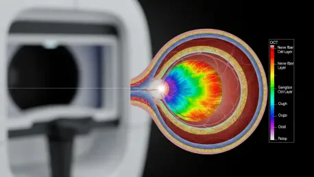
Beyond simple structural imaging, this platform excels at multimodal signal processing, which allows for the creation of enriched feature representations that go far beyond what the human eye can see. Traditional scans often struggle to distinguish between healthy and malignant cells when their refractive indices are similar, but this new tool generates spatially resolved maps of optical attenuation coefficients and speckle contrast metrics. By analyzing how light is absorbed or scattered as it penetrates different layers of tissue, the software can highlight microscopic density variations and depolarization ratios that serve as early indicators of pathological change. These advanced parameters act as a high-definition filter for the human body, significantly enhancing the contrast between different tissue types. For a surgeon, this means having the ability to visualize precise tumor margins that would otherwise be invisible on a standard structural scan, providing a level of intraoperative clarity that was previously reserved for post-operative pathology reports.
Generating Synthetic Data with the Virtual Scanner
A secondary but equally transformative feature of this platform is the Virtual Scanner, an innovative tool designed to solve the chronic shortage of high-quality, labeled datasets in the biomedical imaging sector. In the current environment, obtaining perfectly “ground-truth” data from real human subjects is an incredibly slow and expensive process, often hindered by the natural heterogeneity of biological samples. The Virtual Scanner bypasses these limitations by employing a physics-based simulation engine to generate realistic digital phantoms that mimic the optical behavior of real tissue. Researchers can meticulously control every variable within these synthetic environments, from the density of scatterers to the mechanical elasticity of the simulated sample. This provides a rigorous and repeatable testbed for validating new signal-processing algorithms, ensuring that they are robust enough for clinical use. By providing a controlled space where the “truth” is known with absolute certainty, the platform fosters a more disciplined approach to computational tool development.
The efficacy of this synthetic approach has been validated through rigorous experimental testing involving a wide array of biological models, ranging from murine tumor growth to human brain and endometrium samples. These tests demonstrated that the platform’s multimodal analysis provides a far more comprehensive picture of tissue health than traditional structural imaging could ever achieve on its own. This capability is pushing the medical community closer to the realization of a true “optical biopsy,” a non-invasive procedure that could eventually replace the need for physical tissue removal. By enabling real-time, high-resolution assessments during a surgical procedure, the platform allows for immediate diagnostic feedback, which is crucial for improving patient outcomes. This shift toward non-invasive diagnostics not only reduces the risk of complications associated with traditional biopsies but also accelerates the entire treatment pipeline, allowing doctors to make life-saving decisions while the patient is still on the operating table.
Global Innovation and Technical Integration
To foster a spirit of global collaboration and rapid advancement, the developers launched the SynthOCT 2026 Challenge, an initiative that invites the worldwide research community to refine the generation of digital phantoms. This competition focuses on creating scatterer distributions that not only look like real tissue but also behave like real tissue when subjected to the platform’s Virtual Scanner. Such collaborative efforts are essential for building the massive, physically consistent datasets required to train the next generation of artificial intelligence and foundation models for virtual histology. As machine learning becomes more integrated into healthcare, the quality of training data becomes the primary determinant of success. By crowdsourcing the improvement of these digital phantoms, the consortium is ensuring that future AI tools will be grounded in solid physical principles rather than mere statistical correlations, leading to more accurate and trustworthy diagnostic outcomes in the years ahead.
The long-term impact of this technology was rooted in its ability to blend high-end cloud computing with a simplified user experience, creating a scalable ecosystem for the future of oncology. By abstracting the most difficult computational workflows, the platform opened the door for a much broader range of medical professionals to contribute their expertise to the field of optical diagnostics. The integration of no-code software, physics-based modeling, and multimodal analytics successfully addressed the dual challenges of technical complexity and data scarcity that had long hindered progress. As these tools became more prevalent in clinical settings, they bridged the gap between advanced imaging science and practical bedside care, offering a clear path toward personalized medicine. Stakeholders in the medical technology sector were encouraged to integrate these open-access platforms into their existing diagnostic frameworks to maximize patient benefit. The transition toward precise, real-time guidance established a new standard for intraoperative care, ensuring that imaging technology functioned as a true partner in the healing process.