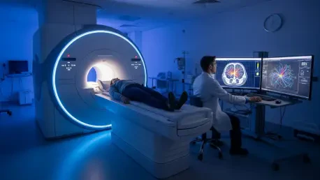

The traditional constraints of medical resonance imaging have long forced a difficult compromise between the depth of biological data collected and the time a patient spends inside a scanner. While the medical community has relied on MRI as a gold standard for decades, the technology has primarily functioned as a sophisticated camera for water molecules, providing clear anatomical structures but leaving the complex chemistry of the brain largely invisible. This structural focus means that critical information regarding metabolic health, neurotransmitter levels, and cellular integrity remains trapped within the signals, often requiring multiple, lengthy sessions to extract even a fraction of the available data. A transformative leap from the University of Illinois Urbana-Champaign has introduced Multiplexed MRI, or MRx, which effectively redefines the role of the scanner from a structural imaging tool into a high-speed molecular analyzer. By shifting the focus to a holistic view of the brain’s physiological landscape, this technology allows for the simultaneous mapping of dozens of biomarkers that were previously too faint or too slow to capture in a clinical setting.
Overcoming Dimensionality With Advanced Physics
The primary technical hurdle in high-dimensional imaging is often described as the curse of dimensionality, where the addition of each new variable typically results in an exponential increase in the time required for data acquisition. To bypass this fundamental limitation, the MRx system utilizes a specialized pulse sequence coupled with a sparse sampling framework that avoids the need to record data points sequentially. Instead of focusing on one chemical signature at a time, the scanner excites and encodes all detectable molecular signals simultaneously across the entire brain volume. This approach dramatically compresses the timeline of the scan, ensuring that the vast array of data required for a comprehensive molecular map can be gathered within a single, continuous session. By optimizing the way the magnetic field interacts with various tissue components, the researchers have managed to preserve signal integrity while moving through the acquisition process at a pace that was previously considered impossible for such complex datasets.
Once the raw data is captured, the challenge shifts to the immense difficulty of distinguishing between overlapping signals that are often ten thousand times weaker than those produced by water protons. Conventional reconstruction software lacks the sensitivity to separate these faint chemical echoes from the background noise, leading to blurred or inaccurate measurements of vital metabolites. The research team addressed this by integrating physics-driven machine learning algorithms directly into the processing pipeline, allowing the system to untangle the complex interference patterns inherent in multiplexed data. These algorithms are trained on the underlying laws of nuclear magnetic resonance, enabling them to predict and isolate the specific signatures of individual biomarkers with high precision. This marriage of physical principles and computational intelligence ensures that the resulting maps are not just images, but quantified data sets that provide a clear and objective window into the chemical and microstructural state of the human brain.
Redefining Efficiency in Clinical Neuroimaging
The performance benchmarks established by this new technology represent a significant departure from existing protocols, which often require patients to remain perfectly still for over an hour to achieve multi-contrast results. In a single 14-minute session, MRx can successfully quantify 22 distinct biomarkers, providing a high-resolution spectrum that covers everything from tissue metabolism to physiological function. This efficiency is particularly critical for patients who may struggle with the claustrophobic environment of a standard MRI machine or those with neurological conditions that make long periods of immobility difficult. By reducing the time burden while simultaneously increasing the volume of data collected, the technology bridges a longstanding gap between high-end scientific research and the practical needs of a busy hospital environment. The ability to generate such a wealth of information in under a quarter of an hour ensures that comprehensive molecular profiling can finally become a routine part of standard neurological care.
Beyond the immediate benefits of speed, the high-resolution nature of MRx data provides a level of detail that surpasses what is currently available through standard clinical sequences. Traditional scans often provide a qualitative “picture” that requires subjective interpretation by a radiologist, but MRx delivers standardized, quantitative measurements of the brain’s functional state. This shift toward quantification means that physicians can compare a patient’s molecular profile against a normative database, identifying subtle deviations that might suggest the presence of a burgeoning pathology. The technology captures a wide array of information including axonal density, myelin integrity, and metabolic concentrations, all within a unified coordinate system. This holistic approach ensures that no single aspect of brain health is viewed in isolation, allowing for a more integrated understanding of how different biological systems interact during the progression of a disease or in response to a specific medical treatment.
Precision Applications in Oncology and Chronic Disease
In the specialized field of neuro-oncology, the precision offered by MRx provides a vital tool for navigating the internal complexity of brain tumors. Standard imaging can effectively locate a mass, but it often fails to describe the heterogeneous environment within the tumor itself, such as the specific areas of high metabolic activity versus regions of fluid buildup. By generating a comprehensive tissue state index, MRx allows clinicians to distinguish between eight different tissue types, including healthy gray matter, white matter, and varying grades of malignant growth like glioblastoma or meningioma. This level of differentiation is essential for the planning of radiation therapy, as it enables oncologists to target the most aggressive parts of a tumor while sparing the surrounding healthy tissue from unnecessary damage. The ability to visualize these boundaries with chemical precision reduces the guesswork often associated with aggressive brain cancers, leading to more tailored and effective surgical and radiological interventions.
The diagnostic advantages of this technology also extend to the long-term management of neurodegenerative and inflammatory conditions like Multiple Sclerosis. One of the most significant challenges in treating MS is determining whether a lesion is currently active or representing chronic, stable damage, a task that typically requires the injection of gadolinium-based contrast agents. MRx eliminates this necessity by directly identifying biomarkers associated with active demyelination and axonal damage through purely non-invasive means. By tracking these molecular markers over time, physicians can predict the likely progression of a lesion and adjust medication strategies before physical symptoms worsen. This proactive approach to disease management is also being explored for epilepsy, stroke, and Alzheimer’s disease, where identifying a molecular fingerprint of dysfunction can lead to much earlier detection. Catching these metabolic shifts before structural changes occur represents a major step forward in the movement toward personalized, data-driven healthcare.
Global Implementation and Next Steps for Imaging
A major factor facilitating the rapid adoption of MRx is its inherent compatibility with the existing infrastructure found in modern diagnostic facilities. Unlike many emerging technologies that require the purchase of multi-million dollar hardware, MRx is primarily a software-driven innovation that can be integrated into the 3 Tesla scanners already ubiquitous in hospitals. This means that a standard imaging center can be upgraded to provide advanced molecular mapping without undergoing a massive capital expenditure or a complete overhaul of their facility. The licensing of this technology for clinical evaluation at centers across the globe marks the beginning of a new era where high-level metabolic data is accessible to the general public. As the software is refined and distributed, the consistency of the data sets produced will allow for a more unified global standard in how brain health is measured and monitored across different populations and various clinical settings.
Looking toward the immediate future, the roadmap for this technology involves expanding the scope of its detection capabilities to include multinuclear imaging. While current versions of MRx focus on proton-based signals, upcoming iterations are expected to track elements such as sodium, phosphorus, and deuterium, which will offer even deeper insights into the body’s energy metabolism and ion balance. This expansion will likely provide new perspectives on how the brain recovers from injury and how it processes nutrients at a cellular level. Clinical teams should prepare for this transition by standardizing their data storage and analysis pipelines to handle the influx of quantitative information. The shift from looking at simple anatomical pictures to analyzing complex chemical maps was established through rigorous testing and is now moving into a phase of broad clinical validation. Moving forward, the focus will remain on refining these algorithms to ensure they provide actionable insights for every patient who enters a scanner.