
Chronic liver disease currently affects millions of individuals globally, yet the diagnostic journey remains fraught with fragmented procedures that often delay critical interventions and treatment. The introduction of the Mindray Hepatus Series marks a definitive shift toward an integrated diagnostic ecosystem where high-resolution imaging and quantitative tissue analysis no longer exist as separate clinical silos. By consolidating these functions into a unified workflow, healthcare providers can now address the complex needs of patients with non-alcoholic fatty liver disease or chronic hepatitis with unprecedented precision. This evolution is not merely a hardware upgrade but a fundamental reimagining of how gastroenterology and hepatology departments operate on a daily basis. Instead of patients moving between multiple rooms for morphological scans and specialized elastography, the entire assessment occurs in one seamless encounter, significantly reducing the diagnostic window while improving the overall quality of care.
Integrating Morphology and Tissue Mechanics
Versatile Platforms: Addressing Clinical Diversity
Focusing on the flagship Hepatus 6, this system provides a robust solution for large hospital environments where high patient throughput and multi-organ diagnostic capabilities are essential. It serves as a comprehensive imaging workstation, capable of detailed evaluations that extend beyond the liver to include the spleen and the broader gastrointestinal tract. This versatility is crucial for identifying secondary complications of portal hypertension or other systemic manifestations of chronic liver issues. By offering superior image quality and specialized tools in a single console, the system allows radiology and GI departments to maximize their resources without sacrificing the depth of the clinical exam. The hardware design emphasizes ergonomic efficiency, ensuring that clinicians can perform numerous scans throughout a shift without the physical fatigue often associated with traditional ultrasound equipment. Such a foundation ensures that the most complex clinical cases receive the necessary attention.
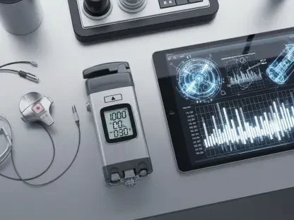
In contrast, the Hepatus 5 addresses the growing need for mobility and bedside care within the modern medical facility through its innovative tablet-based design. This portable model does not compromise on diagnostic power, bringing advanced transient elastography directly to the patient in various settings like intensive care units or remote satellite clinics. For healthcare systems operating across multiple sites, the ability to deploy high-level liver assessment tools outside the central imaging suite is a significant operational advantage. The compact form factor allows for rapid deployment in emergency situations or routine check-ups where space is limited, ensuring that every patient has access to top-tier diagnostic technology regardless of their location. This democratization of care is a hallmark of the series, as it bridges the gap between specialized tertiary centers and community-based practices. Providing this level of accessibility ensures that early-stage liver conditions are detected more frequently.
Visualized Elastography: Precision in Early Detection
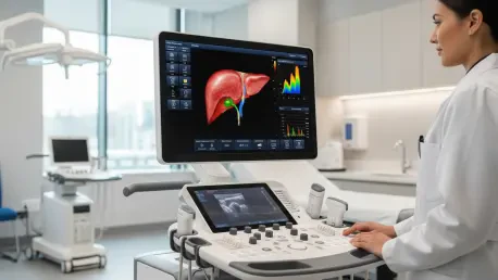
A core technological advancement in these systems is Visualized Transient Elastography, or ViTE, which allows clinicians to see anatomical structures while simultaneously measuring tissue stiffness. This real-time visual guidance is a significant departure from older “blind” elastography methods that relied on surface markers and estimation. By providing a clear view of the target area, ViTE minimizes the risk of measuring non-representative tissues, such as blood vessels or rib shadows, which can often skew results. Research conducted over the current year indicates that this technology significantly enhances the diagnostic accuracy of stiffness measurements, providing a reliable numerical value for liver fibrosis staging. The ability to visualize the region of interest ensures that every data point collected is clinically relevant, reducing the need for repeat scans and providing a higher level of confidence for the medical team. This precision is essential for tracking progress in chronic cases.
Furthermore, the integration of advanced quantification tools helps in the early identification of subtle tissue changes that might otherwise go unnoticed during a standard ultrasound examination. By detecting these changes sooner, medical professionals can implement earlier interventions, potentially avoiding the need for invasive and costly liver biopsies. This non-invasive approach is particularly beneficial for patients who require frequent monitoring, as it eliminates the risks associated with surgical procedures and reduces the overall burden on the healthcare system. The Hepatus Series also includes specialized modules for measuring liver fat content, providing a comprehensive metabolic profile in a single sitting. This multi-parametric approach allows for a more nuanced understanding of a patient’s liver health, facilitating personalized treatment strategies that are tailored to the specific stage and nature of the disease. This shift toward non-invasive quantification is transformative.
Enhancing Reliability Through Intelligent Automation
The AI Co-Pilot: Standardizing Ultrasound Performance
To address the common issue of user variability in ultrasound results, Mindray has integrated AI-driven “co-pilot” features like the Q-Scan Intelligent Acquisition tool. This software automates the collection of valid measurements, ensuring that diagnostic data remains consistent even when different technicians perform the scans over several years. This is a critical development because the longitudinal tracking of liver disease requires data that can be compared accurately across time. The Q-Scan tool uses advanced algorithms to recognize ideal acoustic windows and automatically triggers the measurement when the signal quality is optimal. This reduces the cognitive load on the operator, allowing them to focus more on the patient and less on the technical intricacies of the machine. By standardizing the way data is acquired, the system ensures that changes in a patient’s condition are a result of clinical progression rather than variations in scanning technique or operator experience.
In addition to automated data collection, the system features a Quality Control Index that offers real-time feedback to the operator during the scanning process. By monitoring factors such as probe pressure and patient respiratory motion, the system guides the user to maintain the ideal conditions for a successful scan. This built-in quality assurance democratizes advanced diagnostics, enabling operators with varying levels of experience to achieve professional-grade results. If the probe pressure is too high or the patient’s breathing interferes with the measurement, the system provides an immediate visual alert, allowing for instant correction. This proactive approach to quality control significantly reduces the rate of discarded or inaccurate measurements, which traditionally plague ultrasound-based assessments. Consequently, clinics can maintain a high standard of care without needing a team of highly specialized elastography experts, making advanced liver diagnostics more sustainable.
Strategic Integration: Establishing a New Clinical Standard
The implementation of these advanced diagnostic systems established a new benchmark for hepatic care, as medical facilities successfully integrated quantitative analysis into their daily routines. Healthcare organizations identified that the transition toward automated, AI-assisted workflows was the most effective method for mitigating the risks of operator dependency. It became apparent that the shift from fragmented scanning to unified multi-organ assessments delivered superior clinical insights while optimizing departmental efficiency. Professionals recognized that maintaining a secure, structured data environment was essential for longitudinal disease management and improved patient safety. By adopting these integrated ecosystems, clinics achieved a more proactive stance in identifying metabolic disorders and chronic liver conditions before they reached critical stages. The focus transitioned toward utilizing these high-resolution datasets to refine personalized therapy and enhance diagnostics.
Facilities that embraced the Living Technology software pathway ensured that their diagnostic capabilities remained current without requiring frequent hardware overhauls. This approach allowed clinical teams to access the latest investigative tools and measurement algorithms as soon as they became available, maintaining a cutting-edge environment for patient care. The strategic focus moved toward expanding the use of non-invasive elastography across a wider range of gastrointestinal conditions, reflecting a comprehensive understanding of systemic health. Administrators found that investing in such interconnected platforms reduced the long-term operational costs associated with diagnostic errors and redundant testing. Ultimately, the adoption of this technology facilitated a more collaborative relationship between radiologists, hepatologists, and primary care physicians. This unified front empowered medical professionals to deliver consistent, data-driven outcomes that addressed the complex realities of liver and GI health.