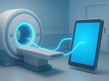

The rapid evolution of medical technology has fundamentally changed how radiologists approach patients with metallic implants. Clinical safety protocols rely on a rigorous verification process known as the identification pyramid, which prioritizes the physical implant card as the definitive source of truth. In the current medical environment, the focus has shifted from simply avoiding these patients to carefully managing the specific risks associated with each unique device to ensure a safe and effective diagnostic outcome. Despite these advancements, a significant gap in safety knowledge persists within the medical community. Research indicates that many hospitals still experience avoidable accidents, often because staff are not fully aware of the most current safety guidelines. This lack of information sometimes leads to diagnostic nihilism, where patients are denied necessary scans for devices that are actually safe, highlighting the urgent need for better education and more standardized protocols in imaging departments across the country. By moving toward a culture of evidence-based safety, the industry ensures that every patient has access to high-quality imaging regardless of their surgical history.
The Mechanics of Magnetic and Thermal Interactions
The primary concern when introducing an implant into the MRI environment involves the immense power of the static magnetic field, which can interact with ferromagnetic materials in ways that are both sudden and violent. If a device contains significant amounts of iron, cobalt, or nickel, the magnetic field exerts a translational force that pulls the object toward the center of the bore, potentially turning a small implant into a dangerous internal projectile. Even if an object is securely anchored in tissue, it may still experience torque, a rotational force that attempts to align the long axis of the implant with the magnetic lines of force. This is particularly critical for sensitive placements, such as intracranial aneurysm clips or ocular implants, where even a microscopic shift could result in catastrophic physiological damage. The intensity of these forces increases as the patient moves closer to the magnet’s center, making the physical positioning of the patient a crucial component of the initial safety assessment. Modern facilities must employ highly trained personnel who understand the spatial gradient of the magnetic field to prevent accidental displacement during the entry and exit phases of the scanning procedure.
Beyond the static field, the active scanning process introduces risks related to radiofrequency pulses and changing magnetic gradients that can lead to significant thermal injury. These radiofrequency pulses can induce electrical currents in metallic leads or long implants, creating what is known as the antenna effect, where the tips of an implant become hot enough to burn surrounding biological tissue. This phenomenon is a major concern for patients with cardiac pacemakers or deep brain stimulators, where the leads act as conductors for the energy delivered by the scanner. Additionally, rapidly switching magnetic gradients can cause certain implants to vibrate, resulting in patient discomfort or the minor migration of the device during the procedure. To manage these risks, technologists must carefully calibrate the scanner to stay within specific energy limits that prevent excessive heating. Understanding these physical interactions is not merely an academic exercise but a practical necessity for ensuring that the benefits of an MRI scan are not outweighed by the potential for internal thermal or mechanical trauma.
Navigating Device Labeling and Modern Safety Metrics
The medical device industry currently categorizes implants as MR Safe, MR Unsafe, or MR Conditional, but these labels require a sophisticated understanding to be applied correctly in a clinical setting. The MR Conditional category is particularly complex because it signifies that the device is only safe under a very specific set of circumstances, such as a maximum static magnetic field strength or a limited spatial gradient. Radiologists must verify that the scanner’s technical specifications and the energy output of the planned sequence match the exact requirements set by the device manufacturer. Any deviation from these conditions, however slight, can lead to serious safety failures or device malfunctions. In practice, this means that a device cleared for a 1.5-Tesla scanner may be entirely unsafe in a 3-Tesla environment, necessitating a fresh review of manufacturer documentation for every individual case. The challenge lies in the sheer volume of new devices entering the market, which requires imaging departments to maintain updated databases and robust communication channels with surgical teams.
To improve the accuracy of safety assessments, the medical field is transitioning toward more precise methods of measuring energy exposure during a scan. Traditionally, energy was measured by the Specific Absorption Rate (SAR), which estimates the amount of radiofrequency energy absorbed by the patient’s body mass. However, experts now prefer using the B1+rms metric, which measures the actual magnetic field delivered by the scanner regardless of the patient’s size or physical composition. This shift allows for more consistent safety checks across different patient populations and ensures that the energy levels used during the scan remain within the safe thresholds established for specific implants. By adopting B1+rms as a primary metric, radiologists can more confidently manage patients with implants that have narrow safety margins. This technical transition represents a move toward a more objective and standardized safety framework, reducing the reliance on generalized estimates and providing a more granular understanding of how electromagnetic energy interacts with the patient’s internal hardware.
Overcoming Image Distortion through Technical Innovation
Even when a scan is deemed physically safe, the presence of metal often creates significant image artifacts that can obscure vital diagnostic information. These distortions typically appear as signal voids or bright “blooming” spots where the metal disrupts the uniformity of the magnetic field, often hiding the very anatomy that the clinician needs to evaluate. In many instances, these artifacts prevent the scanner from properly suppressing fat signals, which is essential for identifying bone marrow edema or inflammation in the soft tissues surrounding a joint replacement. For patients with orthopedic hardware, these distortions can make it nearly impossible to detect early signs of implant loosening or infection. This technical hurdle has historically limited the utility of MRI for postoperative evaluations, but recent advancements in pulse sequence design have begun to address these limitations. By understanding how different metals affect field homogeneity, radiologists can select specific imaging parameters that minimize the visual impact of the implant, thereby restoring the diagnostic value of the procedure.
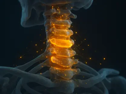
To clear up these images, radiologists utilize a variety of specialized technical strategies and advanced software sequences. One of the most effective methods involves using a lower field strength scanner, such as a 1.5-Tesla unit instead of a 3-Tesla one, which naturally reduces the severity of the metal-induced distortion. Additionally, sophisticated software tools like SEMAC (Slicing Encoding for Metal Artifact Correction) and MAVRIC (Multi-Acquisition Variable Resonance Image Combination) use complex mathematical algorithms to “see through” the interference caused by the metal. These sequences provide much clearer views of the tissue-implant interface, allowing for a detailed assessment of the surrounding bone and ligaments. While these advanced sequences often require longer scan times, the gain in image quality is frequently the difference between a definitive diagnosis and an inconclusive result. The integration of these technologies into standard clinical practice has revolutionized the follow-up care for patients with complex orthopedic or spinal reconstructions, ensuring that metallic hardware is no longer a barrier to high-quality diagnostic imaging.
Strengthening Verification Protocols and AI Integration
The final line of defense in maintaining a safe MRI environment is a strict verification protocol that leaves no room for ambiguity. Professionals are required to confirm the exact make, model, and serial number of an implant by referencing the patient’s device card, operative reports, or specialized manufacturer safety databases. Relying on visual identification from previous X-rays or CT scans is considered a last resort, as many different devices appear nearly identical on plain film but possess vastly different safety profiles under the influence of a powerful magnet. A common misconception in clinical practice is that if a patient has successfully undergone an MRI in the past, they are automatically safe for future procedures. However, safety depends entirely on the specific hardware and software settings of the scanner being used today, which may differ significantly from previous equipment or software versions. Every scan requires a fresh evaluation of the implant’s conditions to account for any changes in the patient’s health or advancements in imaging technology that might have occurred since their last appointment.
Looking toward the future of clinical practice, the integration of artificial intelligence promises to make the screening process both faster and more accurate. AI tools are currently being developed to comb through thousands of pages of electronic medical records to identify forgotten implants that a patient might have overlooked or forgotten during their initial screening interview. These digital assistants can automatically flag potential conflicts and cross-reference them with the latest manufacturer safety updates, providing an extra layer of protection against human error. While human oversight remains the essential centerpiece of any safety program, these automated systems can significantly reduce the administrative burden on technologists and radiologists. By streamlining the identification process, AI-driven tools help ensure that fewer patients are turned away due to missing documentation while simultaneously decreasing the risk of accidental exposure to unsafe magnetic fields. This synergy between human expertise and computational power is defining a new standard for patient safety in the high-tech landscape of modern radiology.
Establishing Resilient Safety Frameworks for the Future
The successful management of medical implants in the MRI suite required a comprehensive overhaul of historical safety frameworks and the adoption of a more proactive clinical stance. Healthcare systems implemented rigorous training programs that prioritized the understanding of electromagnetic physics and device labeling for all frontline staff, from technologists to referring physicians. These initiatives successfully bridged the knowledge gap that previously led to unnecessary scan cancellations, ensuring that patients received timely diagnostic interventions. By formalizing the use of the identification pyramid and prioritizing physical documentation over anecdotal history, facilities established a reliable baseline for risk assessment. This move toward standardized verification protocols became the foundation for a culture where safety was integrated into every step of the patient journey, from the initial consultation to the final imaging report. The industry recognized that the complexity of modern implants demanded a departure from the “one-size-fits-all” approach, leading to more personalized and precise safety evaluations.
Moving forward, the focus turned toward the seamless integration of digital tools and cross-departmental collaboration to maintain these high safety standards. The deployment of AI-enhanced screening software allowed for the rapid cross-referencing of surgical histories with manufacturer safety databases, significantly reducing the potential for human oversight. Furthermore, the clinical community emphasized the importance of detailed operative records, urging surgeons to provide clear, accessible information regarding the MR status of every implanted device. This collaborative effort ensured that the responsibility for safety was shared across the entire continuum of care rather than falling solely on the imaging department. As technology continued to advance from 2026 to 2028 and beyond, these established protocols provided a flexible yet sturdy structure for incorporating new materials and imaging techniques. By fostering an environment of continuous learning and technical adaptation, the medical field successfully transformed the challenge of metallic implants into a manageable aspect of routine diagnostic practice, ultimately improving patient outcomes through safer and more accurate imaging.