
The precision of preoperative management for early-stage cervical cancer is currently undergoing a transformative shift as clinicians look beyond traditional imaging capabilities. At the heart of this evolution is the clinical necessity to identify lymphovascular space invasion (LVSI), a microscopic marker that often dictates the entire course of postsurgical treatment. Because the presence of tumor cells within the blood or lymphatic vessels significantly increases the risk of recurrence, its detection is paramount for determining whether a patient requires aggressive adjuvant pelvic radiotherapy. Machine learning architectures, such as Extreme Gradient Boosting, are now being paired with AI-enhanced imaging to refine preoperative risk stratification. This integration allows for a much more nuanced understanding of the tumor environment before a single incision is made, enabling surgeons to tailor their approach to the specific biological signature of the disease. By addressing the limitations of standard imaging, medical teams are now able to provide a higher standard of personalized care that was previously unattainable through conventional diagnostic means.
Advanced Methodology and Technical Framework
Research Cohort and Scanning Protocols
The development of these advanced predictive models relied on a meticulously curated retrospective analysis involving 117 patients diagnosed with cervical cancer, specifically ranging from FIGO stages IB to IIB. This cohort was selected from a substantial institutional database of nearly 1,400 records to ensure that only the most complete and high-quality histopathological and imaging data were used for model training. Within this refined group, 45 patients were histologically confirmed to be positive for lymphovascular space invasion, while 72 served as the negative control group. This clear categorization allowed the machine learning algorithms to learn the subtle differences between invasive and non-invasive tumor profiles. The use of such a well-defined cohort ensures that the resulting AI models are grounded in verified clinical outcomes, which is a fundamental requirement for any diagnostic tool intended for use in high-stakes oncological decision-making and surgical planning.
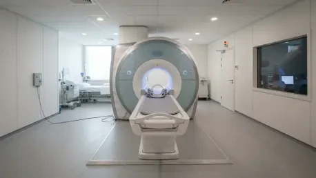
To capture the high-fidelity data required for deep learning, the researchers utilized state-of-the-art Siemens VIDA 3.0-Tesla scanners, which offer superior signal intensity and spatial clarity compared to older imaging systems. The scanning protocol was designed to be comprehensive, incorporating multiparametric sequences such as axial T1-weighted imaging, axial T2-weighted imaging with spectral attenuated inversion recovery (T2-SPAIR), and diffusion-weighted imaging (DWI) with multiple b-values. These sequences provided a rich, multidimensional view of the pelvic anatomy, focusing on the specific textures and water diffusion patterns within the tumor tissue. By collecting this diverse array of imaging data, the researchers provided the AI framework with a robust foundation of information, allowing it to analyze the tumor from various physiological perspectives. This multi-sequence approach is critical for capturing the full spectrum of tumor heterogeneity that standard single-sequence scans often overlook.
The Power of Super-Resolution GANs
The most significant technical breakthrough in this research involves the application of a three-dimensional Super-Resolution Generative Adversarial Network (SRGAN). This specific deep learning architecture is designed to overcome the physical limitations of MRI hardware by mathematically reconstructing high-resolution images from standard-resolution inputs. Unlike traditional interpolation methods that simply stretch pixels and create a blurred effect, the SRGAN uses a generative process to intelligently fill in missing anatomical details based on learned patterns. This resulted in a fourfold spatial upsampling, effectively sharpening the voxel dimensions to a degree that reveals microscopic details of the tumor boundary. For the T2-SPAIR sequences, the resolution was enhanced from 0.781 mm to an impressive 0.195 mm, providing a level of clarity that was previously impossible to achieve without significantly extending the patient’s time inside the scanner.
To train the SRGAN effectively without the need for matched high-resolution medical scans, the team implemented a specialized “frozen inference” strategy. This technique involved taking a model that had been pretrained on vast datasets of high-quality natural images and then applying its learned structural priors to the medical imaging domain. By leveraging these existing patterns of edge definition and texture, the model was able to enhance the medical scans with remarkable accuracy. The process allowed for the creation of high-fidelity images that maintain the original signal integrity while suppressing background noise. This post-processing “upgrade” is particularly valuable in a clinical setting because it improves diagnostic accuracy without requiring any changes to the existing hardware or patient protocols. This makes the technology highly scalable and easily integrated into modern radiology departments that already utilize high-field MRI systems.
Radiomics Workflow and Machine Learning Analysis
Quantitative Feature Extraction and Selection
The conversion of these enhanced images into actionable data was achieved through a sophisticated radiomics pipeline. An abdominal radiologist with over six years of specialized experience performed manual segmentations of the tumor regions of interest using 3D Slicer software. This manual process ensured that the AI was only analyzing the relevant cancerous tissue, effectively filtering out healthy surrounding structures. From these segmented regions, the pipeline extracted a staggering 1,427 quantitative features per imaging sequence. These features ranged from basic morphological measurements like shape and volume to complex high-order texture descriptors that capture the mathematical randomness and uniformity of the tumor’s internal structure. This high-dimensional data provides a “digital biopsy” of the cancer, offering insights into its aggressive nature that the human eye cannot detect during a routine visual review of the scans.
To ensure that only the most reliable and predictive data points were used, the researchers conducted a rigorous feature selection process. This involved a stability analysis where features were tested for reproducibility; any descriptors that showed significant variation over multiple measurements were discarded. The remaining 1,715 stable features were then processed using Least Absolute Shrinkage and Selection Operator (LASSO) regression and Spearman correlation analysis. This statistical filtering narrowed the vast dataset down to a core set of highly predictive variables that directly correlated with the presence of lymphovascular space invasion. By reducing the dimensionality of the data, the researchers were able to prevent the machine learning models from overfitting, ensuring that the final algorithms would be effective when applied to new, unseen patient data. This refined approach to feature selection is what allows the model to maintain high accuracy across different patient demographics.
Evaluating Predictive Model Architectures
The research team evaluated six different machine learning architectures to identify which mathematical framework could best interpret the complex radiomic data. These included Extreme Gradient Boosting (XGBoost), Light Gradient Boosting Machine (LightGBM), support vector machines (SVM), and logistic regression, among others. Each model was rigorously tested on the validation cohort to determine its sensitivity and specificity in detecting LVSI. Throughout the testing phase, the XGBoost model consistently outperformed the other classifiers, demonstrating a superior ability to handle the nonlinear relationships found within the multi-sequence radiomic data. The model’s success is attributed to its gradient boosting framework, which iteratively corrects errors and optimizes the decision-making process. This makes it an ideal choice for the high-stakes environment of oncological diagnostics where accuracy and reliability are the most critical metrics.
One of the most striking findings during the model evaluation phase was the complete dominance of radiomic data over traditional clinical variables. When the researchers attempted to include factors such as patient age, tumor diameter, and FIGO stage into the final model, the LASSO selection process actually excluded them. This indicated that the high-dimensional information captured by the super-resolution radiomics already contained all the necessary predictive variance. The AI found that the mathematical textures within the tumor were much more indicative of vascular invasion than the macroscopic characteristics used in standard staging. This shift away from traditional metrics highlights the potential for AI to redefine how risk is assessed in cervical cancer patients. By relying on objective, data-driven markers rather than subjective clinical observations, the medical community can move toward a more standardized and accurate method of preoperative risk assessment.
Clinical Results and Future Implications
Significant Gains in Diagnostic Performance
The statistical results of the study provided clear evidence that super-resolution enhancement is a game-changer for diagnostic accuracy. In the internal validation cohort, the model utilizing super-resolution diffusion-weighted imaging (SR-DWI) achieved an area under the receiver operating characteristic curve (AUC) of 0.864. This was a substantial leap over the 0.727 AUC produced by the original-resolution imaging. However, the most powerful results were found in the multi-sequence fusion model, which combined the best features from both DWI and T2-SPAIR sequences. This ultimate model reached an AUC of 0.934, indicating an exceptionally high level of precision. Such a significant improvement proves that the additional spatial detail provided by the SRGAN allows the AI to “see” patterns of invasion that are completely invisible on standard MRI scans, providing a much clearer picture of the patient’s risk profile.
Beyond the raw performance numbers, the consistency of the improvements across all six machine learning models was particularly noteworthy. Every single algorithm tested, from the complex neural networks to the simpler linear models, showed a marked increase in predictive power when fed with super-resolution data rather than original scans. For example, the LightGBM model saw its performance jump from a mediocre AUC to a highly reliable one simply by switching the input data type. This consistency suggests that the benefit of super-resolution is not dependent on a specific type of code, but is instead a fundamental improvement in the quality of the underlying medical information. This finding is critical for the widespread adoption of AI in radiology, as it demonstrates that enhancing image quality is a universal way to boost the performance of any digital diagnostic tool currently in development.
Defining the New Standard of Care
The investigation at The First People’s Hospital of Guangyuan successfully demonstrated that 3D super-resolution reconstruction served as more than just a visual aid; it acted as a vital diagnostic enhancer. By mathematically refining the spatial resolution of conventional MRI, the researchers unlocked a deeper layer of previously inaccessible data regarding tumor heterogeneity. The high performance of the SR-fusion model suggested that AI-enhanced radiomics was prepared to become a standard component of the preoperative workup for cervical cancer. The study proved that these models provided a significant net benefit in clinical decision-making, as verified by Decision Curve Analysis and Net Reclassification Improvement metrics. These tools confirmed that the AI could reliably categorize patients into high-risk or low-risk groups, which allowed for the creation of highly personalized and effective treatment strategies that minimized unnecessary surgical interventions while targeting aggressive disease more effectively.
Moving forward, the clinical implementation of these AI-enhanced workflows will require the establishment of multicenter trials to validate the models across more diverse patient populations and different imaging hardware. Future efforts should focus on integrating these high-resolution algorithms directly into the radiology workstation, allowing for real-time risk assessment during the initial diagnostic phase. Additionally, expanding the scope of the AI to include other biological markers, such as human papillomavirus status and specific serum tumor antigens, could result in even more comprehensive predictive tools. As these technologies mature, they will likely redefine the standard of care in gynecological oncology, shifting the focus toward a data-driven approach that prioritizes precision and patient safety. The foundation established by this research has already paved the way for a new era of oncological imaging where the microscopic details of a tumor are no longer hidden from the clinical team.