
The determination of lymph node involvement remains the single most critical factor in deciding the optimal therapeutic path for individuals diagnosed with early-stage cervical cancer. Traditionally, high-cost imaging modalities like Magnetic Resonance Imaging and Positron Emission Tomography have served as the gold standards for identifying metastatic spread within the pelvic region. However, the complexity and expense associated with these technologies often create barriers to timely care, especially in resource-limited clinical settings where specialized equipment is not readily available. Recent clinical data suggest that high-resolution pelvic ultrasound, when performed by experienced sonographers, provides diagnostic accuracy comparable to these advanced cross-sectional imaging techniques. This shift in perspective is prompting a reevaluation of standard preoperative protocols, potentially allowing for more streamlined and cost-effective patient management strategies that do not sacrifice the precision required for oncological decision-making.
Comparative Efficacy of Diagnostic Modalities
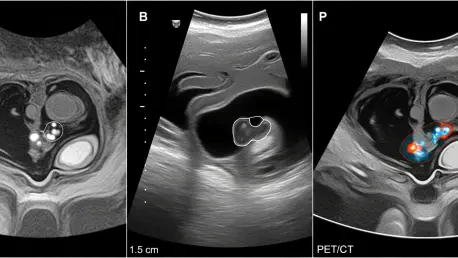
Performance Metrics: Evaluating Nodal Detection Accuracy
Clinical investigations have recently demonstrated that transvaginal and transrectal ultrasound can identify pelvic lymph node metastasis with a level of precision that mirrors the performance of traditional MRI and PET/CT scans. In a comprehensive prospective study involving hundreds of patients, the sensitivity and specificity of ultrasound were found to be statistically non-inferior to its more expensive counterparts. This is particularly significant because ultrasound allows for a real-time, dynamic assessment of the pelvic anatomy, providing clinicians with immediate insights into the morphology and vascularity of suspicious nodes. While MRI remains excellent for assessing the primary tumor size and local extension, ultrasound has proven to be an agile tool for nodal staging. By utilizing high-frequency probes, specialists can detect subtle changes in the architecture of the nodes, such as the loss of the fatty hilum or the presence of abnormal blood flow patterns, which are indicative of malignancy.
Patient Safety: Minimizing Exposure to Ionizing Radiation
The integration of ultrasound into the initial staging process offers a non-invasive alternative that avoids the use of ionizing radiation and contrast agents required for PET/CT and certain MRI protocols. This advantage is particularly relevant for patients with renal impairment or those who experience claustrophobia during long scanning sessions in confined bores. The comparative data highlights that for the detection of pelvic lymph nodes, ultrasound achieved a negative predictive value that ensures clinicians can confidently proceed with surgical interventions when no suspicious nodes are visualized. This level of reliability bridges the gap between affordable diagnostic tools and high-end radiological assessments. As healthcare systems strive to optimize patient throughput, the ability to perform an accurate nodal assessment in an outpatient setting without the need for complex scheduling or specialized radioactive tracers represents a significant leap forward.
Strategic Integration in Clinical Pathways
Implementation: Training and Resource Optimization
The successful adoption of ultrasound as a primary staging tool depends heavily on the standardization of scanning techniques and the rigorous training of medical professionals. Expert sonography requires a deep understanding of pelvic anatomy and the specific criteria used to differentiate between reactive and metastatic lymph nodes. To facilitate this transition, several international oncology societies have begun developing standardized reporting systems and quality assurance programs aimed at ensuring consistency across different institutions. By investing in the specialized training of radiologists and gynecologic oncologists, healthcare facilities can maximize the utility of their existing ultrasound equipment. This approach not only enhances the diagnostic capabilities of the clinical team but also fosters a more multidisciplinary environment where imaging findings are directly correlated with surgical and pathological outcomes.
Future Protocols: Sustainable Diagnostic Practices
Stakeholders in the medical community recognized that expanding the use of pelvic ultrasound significantly lowered the financial burden on patients while maintaining high standards of oncological care. It was observed that hospitals which prioritized ultrasound training saw a reduction in the time between diagnosis and the commencement of treatment, leading to better resource allocation. To capitalize on these findings, clinicians implemented robust internal auditing processes to track the accuracy of their ultrasound assessments against final histopathology reports. Looking ahead, the focus shifted toward integrating artificial intelligence to assist in the identification of metastatic patterns, further reducing operator dependence. Medical boards recommended that specialized training modules become a mandatory component of oncology fellowships to ensure widespread competency. By adopting these measures, the healthcare industry successfully balanced the need for precision medicine with the necessity of sustainable practices.