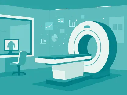

While perfusion measures help predict the recurrence of strokes within 90 days, their current predictive power remains moderate with an AUC of 0.70. Recent clinical research has pivoted toward a multi-modal approach to overcome these limitations, utilizing a retrospective analysis of 353 patients to refine how medical professionals forecast recovery. By integrating clinical bedside scores with multiparametric magnetic resonance imaging taken within the critical twenty-four-hour window of symptom onset, researchers achieved a mean door-to-imaging time of just twenty-seven minutes. This rapid diagnostic pipeline allowed for the evaluation of diffusion-weighted imaging alongside perfusion parameters and susceptibility-weighted imaging to identify reliable indicators for long-term disability. The primary goal was to move beyond simple observation and toward a robust system capable of predicting mortality and hemorrhagic transformation with high precision. This intersection of radiology and clinical assessment marks a significant step in acute stroke management.
Predictive Biomarkers for Mortality and Functional Recovery
The research highlights a profound correlation between specific imaging biomarkers and patient outcomes, particularly when analyzing high National Institutes of Health Stroke Scale scores. Patients presenting with larger diffusion-weighted imaging lesion volumes and prolonged mean transit times consistently demonstrated increased mortality rates and more severe long-term disability. Conversely, the study found that lower values in cerebral blood flow and cerebral blood volume acted as inverse predictors of recovery, signaling a higher risk for neurological decline. One of the most significant findings was the achievement of a ninety-five percent predictive accuracy for mortality by combining specific clinical and radiological thresholds. Specifically, an NIHSS score of at least 10.5, when paired with a lesion volume of 52.5 milliliters or greater and a cerebral blood flow threshold of 20.5 or less, provided a highly reliable prognostic model. This granular data allows clinicians to categorize patient risk profiles with unprecedented clarity during the initial phase.
Beyond mortality, identifying the risk of hemorrhagic transformation remains a primary concern for neurologists managing acute ischemic stroke cases. This frequent and dangerous complication often dictates the safety and efficacy of reperfusion therapies. By applying a threshold of 8.5 for both the NIHSS score and the mean transit time, the study reached a ninety-four percent accuracy rate within the specific cohort observed. This level of precision suggests that the physiological state of the brain tissue, as measured by transit time, is as critical as the external clinical presentation. While clinical comorbidities like hypertension, diabetes, and a history of previous strokes were reaffirmed as significant predictors of poor long-term outcomes, the addition of specific hemodynamic measurements provides a more dynamic view of the patient condition. These findings emphasize the necessity of quantifying physiological parameters rather than relying solely on observational scales to anticipate secondary complications that could derail the recovery process significantly.
Refining Future Diagnostic Protocols and Clinical Implementation
Despite the high accuracy rates reported in this study, researchers have highlighted a critical caveat regarding the general application of these findings. Because the results represent internal discrimination within a specific retrospective cohort, the performance estimates require further testing against diverse, independent populations. The identified thresholds for lesion volume and blood flow remain investigational until validated by broader prospective studies. However, the study strongly supports the potential of radiomics and machine learning to refine stroke prognosis by processing these complex variables simultaneously. Integrating artificial intelligence could soon allow for real-time risk assessment, synthesizing hundreds of radiological features that the human eye might overlook. As the field moves from 2026 to 2028, the focus will likely shift toward automating these measurements to ensure consistency across different medical centers. The current research provides the necessary baseline for these upcoming digital tools, ensuring they are grounded in verifiable physiological data rather than purely theoretical models.
Future advancements in stroke care demanded a shift toward standardized imaging protocols that prioritize rapid and comprehensive data acquisition. Medical institutions moved to integrate these multi-parametric MRI measurements into routine emergency workflows to ensure that prognostic insights were available within the first hour of admission. The focus shifted to developing specialized software that could automatically calculate lesion volumes and perfusion thresholds, reducing the cognitive load on radiologists during high-pressure scenarios. Validation in diverse and prospective cohorts became the primary objective for researchers seeking to bridge the gap between retrospective discovery and bedside application. By establishing these precise diagnostic markers, the medical community laid the groundwork for personalized treatment plans that accounted for individual hemodynamic profiles. Ultimately, the integration of clinical scores with quantitative imaging features provided a clearer path forward for managing complex ischemic events. This proactive approach ensured that high-risk patients were identified earlier, allowing for more aggressive monitoring and targeted interventions.