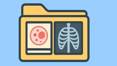

Michigan Medicine became the first United States healthcare system to successfully implement a DICOM-based digital pathology PACS within a broader enterprise imaging network. This milestone signifies a major shift where pathology is no longer treated as an isolated laboratory function but as a critical component of a comprehensive medical imaging strategy. By moving away from traditional glass slides and adopting a fully digital framework, healthcare institutions are dismantling the clinical silos that have long hindered efficient data sharing and diagnostic speed. This transformation allows pathology to follow the path previously blazed by radiology, yet it does so with the benefit of modern software architectures and high-capacity storage solutions. The transition enables a seamless flow of information that connects pathologists directly with the broader clinical team, ensuring that high-resolution tissue images are just as accessible as any radiological scan or clinical note within a patient’s record.
Overcoming Proprietary Barriers With DICOM Standards
The core of this digital evolution lies in the concept of technological leapfrogging, where pathology departments benefit from the maturation of communication standards. While early digital transitions in other medical fields were plagued by proprietary file formats that locked data into specific vendor platforms, current implementations utilize the Digital Imaging and Communications in Medicine (DICOM) standard. This maturity allows healthcare systems to bypass the technical bottlenecks of the past and adopt a universal language for medical images. By prioritizing non-proprietary formats, institutions ensure that their whole slide imaging data remains interoperable across various software platforms and diagnostic viewers. This standardized approach is essential for long-term sustainability, as it prevents the expensive and complex data migrations that often occur when software needs upgrading or vendor contracts change. Consequently, the adoption of DICOM serves as the foundational architecture for a modern system.
Beyond the immediate interoperability, the use of Vendor Neutral Archives (VNA) provides a scalable and cost-effective solution for storing the massive data volumes generated by high-resolution digital slides. Unlike traditional laboratory-specific storage that exists in a vacuum, a VNA allows pathology data to be housed within the same enterprise-level storage environment used by radiology, cardiology, and other specialties. This centralization simplifies data management and security protocols while significantly reducing the overhead associated with maintaining separate hardware for each department. Hospitals can now leverage their existing high-speed network infrastructures to handle the heavy lifting of digital pathology without needing to reinvent the wheel for every new clinical application. This architectural efficiency not only supports the current diagnostic needs but also prepares the institution for future growth as scanning volumes increase. The result is a robust, resilient system that places pathology at the center of the informatics ecosystem.
Creating a Unified Clinical Record for Multidisciplinary Care
Integrating pathology into an enterprise network creates a truly Unified Clinical Record, where the visual evidence of a patient’s condition is no longer fragmented across different systems. When pathology data coexists with images from dermatology, radiology, and other clinical specialties, it provides a comprehensive view of the patient’s journey through the healthcare system. Any authorized clinician can access the full history of diagnostic images directly from the Electronic Health Record (EHR), eliminating the need for manual requests or physical slide transfers. This level of accessibility is particularly vital for chronic disease management and oncological cases, where multiple specialists must collaborate on a single diagnosis. By breaking down these data silos, healthcare organizations foster an environment of transparency and speed, ensuring that clinicians have all the relevant diagnostic tools at their fingertips. This streamlined access reduces the risk of diagnostic errors that can occur when critical information remains buried in an isolated department.
From a financial perspective, the move toward enterprise integration addresses the economic realities of modern hospital budgets by avoiding the “false economy” of siloed systems. While a standalone pathology PACS might appear cheaper during the initial planning phases, it often creates hidden costs that manifest during later integration attempts. By piggybacking on the established IT infrastructure of more mature digital departments, pathology can utilize existing security protocols, backup systems, and storage arrays. This shared resource model reduces the total cost of ownership over a ten-year horizon and ensures that the pathology department does not become a technical island. Furthermore, centralizing the management of digital imaging allows for better negotiation with vendors and more efficient use of technical support staff. Instead of managing multiple disparate contracts, the hospital can focus on a unified strategy that supports the entire enterprise. This strategic alignment ensures that financial investments deliver maximum value.
Advancing Diagnostic Workflows and AI Implementation
Transitioning to a digital environment is not merely about replacing microscopes with monitors; it is about a fundamental redesign of the diagnostic process to improve efficiency and accuracy. Modern digital viewers allow pathologists to display multiple tissue stains from the same patient side by side, facilitating a more comprehensive comparison that is physically impossible with traditional glass slides. This digital workflow also revolutionizes multidisciplinary collaboration, particularly during tumor board meetings where specialists from different departments must review a case simultaneously. In an integrated system, pathologists and radiologists can view tissue samples and radiological scans concurrently within a shared digital space, fostering real-time communication. This instant availability of data accelerates the decision-making process for complex treatment plans, ensuring that patients receive the most accurate diagnosis in the shortest possible time. The digital interface acts as a bridge between medical specialties.
The most significant technological driver for this shift is the emergence of Artificial Intelligence (AI) as a transformative tool in diagnostic medicine. AI algorithms require vast quantities of structured, high-quality data to function effectively, and housing these images within a standardized enterprise architecture provides the necessary training ground. Specifically, multimodal AI platforms, which analyze combined data from radiology and pathology, represent the next frontier in precision medicine. By having both types of diagnostic images stored in a compatible format within the same network, institutions enable AI to analyze the full picture of a patient’s health. This capability allows for the identification of subtle patterns that might be missed by the human eye alone, potentially leading to earlier detection of diseases and more personalized treatment strategies. As the volume of complex cases continues to rise, the integration of AI becomes essential for maintaining high standards of care while managing workloads.
Establishing Robust Governance and Institutional Leadership
Successful implementation of a digital pathology system depends heavily on institutional governance rather than just the selection of advanced hardware or software. A rigorous planning framework must include multi-disciplinary subcommittees that focus on mapping clinical workflows, ensuring interoperability, and providing operational support. It is imperative that pathologists have a primary seat at the table during the platform design phase to ensure the system meets the specific needs of their daily practice. If pathology is not an active participant in these early stages, the department risks being forced to use a platform that was optimized for radiology or other specialties with different requirements. Mapping the journey of a tissue sample from the initial scan to the final diagnostic sign-out ensures that the technology facilitates a smoother experience for the physician. Proper governance also involves establishing clear policies for data access, security, and long-term retention to protect patient privacy while maximizing utility.
Effective leadership is the final piece of the puzzle, requiring a dedicated physician champion who can bridge the communication gap between clinical staff and information technology departments. This individual must be fluent in both medical practice and IT infrastructure to translate clinical needs into technical requirements effectively. Alongside this champion, sustained executive sponsorship from the hospital’s C-suite is vital to navigate the financial complexities and political challenges associated with a project of this scale. Leaders must prioritize the desired future state of the diagnostic workflow rather than simply trying to replicate old analog habits in a digital format. By focusing on the broader strategic goals of the enterprise, healthcare executives can ensure that the transition to digital pathology remains a top priority even during challenging implementation phases. This top-down support provides the necessary resources and organizational alignment to overcome technical hurdles and any potential institutional resistance to the new model.
Leveraging Digital Archives for Modern Research and Trials
The integration of digital pathology into the enterprise network served as a powerful engine for clinical research and academic advancement. Because the data was stored in standardized DICOM formats, it was immediately classified as research-grade, allowing analysts to query the archives using specific diagnostic codes or pathology tags. This standardization eliminated the need for manual data conversion and simplified the process of preparing high-quality datasets for international clinical trials. Automated anonymization profiles ensured that patient confidentiality was maintained without hindering the accessibility of vital diagnostic information for long-term studies. By viewing pathology as a data service rather than a simple lab function, healthcare systems created a sustainable digital ecosystem that improved patient outcomes and streamlined administrative operations. The system allowed researchers to pull vast amounts of data without compromising the performance of clinical diagnostic tools used daily.
Moving forward, institutions should prioritize the recruitment of IT-fluent medical professionals and invest in scalable infrastructure to fully capitalize on these digital assets. The transition demonstrated that success relied not just on software, but on the cultural shift toward open data and cross-departmental collaboration. Healthcare providers were encouraged to audit their existing imaging assets and consolidate them into a single enterprise viewer to maximize the utility of the Unified Clinical Record. The ultimate goal remained the delivery of the right image to the right clinician at the right time, fostering a environment where precision medicine became the standard rather than the exception. By adopting this blueprint, other healthcare systems can replicate the success of early pioneers, ensuring that their diagnostic capabilities remain at the cutting edge of modern medicine. The future of imaging now depends on the seamless integration of every visual data point into a coherent and actionable narrative.