
Medical practitioners have long struggled with the cognitive burden of interpreting two-dimensional slices of the human body to perform complex three-dimensional life-saving interventions in real-time environments. Traditionally, this process, known as mental tomography, requires an incredible amount of focus and years of experience to master effectively. However, a new breakthrough from researchers at the Massachusetts Institute of Technology has introduced a system called Augmented Real-time Volumetric Imaging in Ultrasound, or AR-VIU. This technology essentially provides clinicians with a form of X-ray vision by superimposing high-resolution, three-dimensional digital models of internal tissues directly onto the patient’s body. By shifting the work of spatial reconstruction from the human brain to a sophisticated digital interface, this innovation promises to minimize the potential for human error during diagnostic procedures. This transition toward intuitive, visual-first medical imaging represents a significant shift in how healthcare providers interact with patient anatomy, moving away from flat screens and toward an immersive and highly accurate anatomical reality that was once considered impossible.
Engineering the New Visual Frontier: Technical Specifications
Breakthrough Hardware: The Evolution of Ultrasound Probes
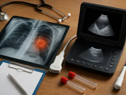
The engineering foundation of the AR-VIU system relies on a specialized ultrasound probe that is roughly the size of a deck of cards, making it remarkably portable compared to traditional devices. Unlike conventional 3D ultrasound equipment, which is often prohibitively bulky and expensive, this new device utilizes a unique empty square array of sensors to gather data. This design allows the probe to capture high-quality volumetric data while using significantly fewer components, which helps in lowering manufacturing costs and reducing overall power consumption. By optimizing the sensor placement, the research team managed to maintain high image fidelity without the need for the massive data processing power usually required for 3D rendering. Because the hardware is more efficient and lightweight, it becomes much easier for medical staff to transport the system between different clinical environments, such as emergency rooms, intensive care units, and even remote field clinics. This accessibility ensures that advanced imaging is not just a luxury for specialized surgical suites.
Real-Time Rendering: Merging Data with Immersive Vision
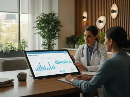
On the software side, the AR-VIU system processes raw data through a high-performance graphics engine that is more commonly found in high-end video games than in a hospital setting. This engine is capable of converting raw ultrasound signals into a direct 3D digital model that preserves all the original spatial relationships and anatomical details. When a healthcare provider wears a compatible augmented reality headset, this digital model is perfectly aligned with the physical body of the patient through sophisticated tracking algorithms. This setup allows the wearer to look directly at the patient and see through the skin, observing internal organs, blood vessels, and tissues from various angles simply by moving their head or changing their physical position in the room. This seamless integration of digital and physical space provides a level of depth perception that 2D screens cannot replicate. By allowing doctors to see the internal environment in its natural context, the system drastically reduces the time needed to identify anomalies or plan a complex surgical approach.
Experimental Success: Validating System Performance
Comparative Analysis: Evaluating Accuracy and Speed
To rigorously confirm that the system actually improves medical performance, the MIT research team conducted a comprehensive series of tests involving both seasoned medical professionals and total novices. Participants were tasked with finding specific objects that were hidden inside opaque gelatin blocks, which were designed to simulate the density and visual resistance of human flesh. They were also asked to mark precise locations on these materials to mimic the high-pressure environment of a needle biopsy or the placement of a surgical tool. These tasks were specifically selected because even a slight deviation in placement can lead to significant clinical complications in a real-world scenario. During these trials, researchers monitored the speed of task completion and the accuracy of the target identification. The goal was to determine if the 3D visual context provided by the AR-VIU system could outperform the traditional methods that doctors have relied on for decades. The experiments provided a wealth of data regarding the usability and reliability of the augmented reality interface.
Skill Acquisition: The Impact on Novice Training
The results of these comparative experiments revealed a massive improvement in both accuracy and speed for every participant, but the impact on beginners was particularly noteworthy. Under traditional 2D imaging conditions, the experts naturally performed much better than the novices, as their years of training allowed them to interpret flat images with higher proficiency. However, when the AR-VIU system was introduced, the performance gap between these two groups almost completely disappeared. Novices were suddenly able to locate and identify hidden objects with nearly the same level of precision and confidence as the seasoned experts. This finding suggests that the 3D visual context provided by augmented reality makes ultrasound technology significantly easier to master, potentially shortening the many years of intensive training usually required to achieve clinical competency. This democratization of skill could allow more medical staff to perform essential imaging tasks without sacrificing the safety of the patient. Moreover, the increased speed of these procedures could lead to higher patient turnover.
Implementation Strategy: Clinical Safety and Precision
Procedural Enhancement: Precision in Invasive Care
The potential clinical benefits of this technology are vast, particularly for invasive procedures such as catheter placements, spinal injections, or delicate biopsies. By seeing the 3D position of a needle relative to a patient’s internal anatomy in real-time, healthcare providers can operate with much greater confidence and a significantly lower risk of accidental tissue damage. While some veteran doctors initially expressed a preference for the traditional 2D methods they had practiced for decades, they quickly acknowledged that the AR-VIU system would be invaluable for high-stakes tasks. For instance, the system showed remarkable potential in monitoring the moving walls of a heart during complex cardiac examinations, where spatial orientation is critical for a correct diagnosis. The ability to see the heart beating in 3D while simultaneously observing the exterior of the patient’s chest provides a comprehensive view of the patient’s condition. This dual perspective ensures that doctors do not lose sight of the broader clinical context while focusing on a specific internal organ or pathological structure.
Future Considerations: The Roadmap for Hospital Use
Researchers focused their final developmental efforts on refining the system to make it even more effective for daily hospital workflows and high-volume clinical use. They prioritized boosting the image resolution to capture even finer biological details, such as small vascular networks and early-stage lesions. The team established a roadmap for more extensive clinical trials, which aimed to ensure the system remained robust across diverse real-world medical environments. Medical institutions were encouraged to evaluate their current imaging infrastructure and consider the long-term benefits of integrating volumetric AR tools into their training programs. This proactive approach helped bridge the gap between experimental laboratory success and practical bedside application. As the technology matured, it transformed into a standard tool within the healthcare industry, making medical imaging more accessible and less time-consuming for everyone involved. Ultimately, the successful implementation of this system provided a clearer path forward for patient safety, proving that the fusion of digital visualization and physical medicine was no longer a theoretical pursuit but a tangible reality.