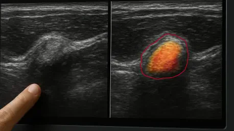

Traditional imaging methods like DECT and MRI offer high accuracy for gout diagnosis but remain limited by their high cost and lack of portability. In the management of advanced gout, the presence of tophi—dense accumulations of monosodium urate crystals—serves as a definitive marker of disease progression and therapeutic response. For years, clinicians have struggled to objectively quantify these masses during routine visits. While physical palpation provides a rough estimate of size, it lacks the precision required to detect subtle changes in volume that indicate whether a specific medication regimen is working. Musculoskeletal ultrasound has emerged as the most practical tool for this purpose, offering a non-invasive way to visualize crystals at a fraction of the cost of advanced scans. However, the reliance on manual segmentation by an operator introduces significant variability, making it difficult to achieve the standardization needed for high-quality clinical care in 2026.
Data Curation: Foundation of the AI Training Pipeline
The success of any medical artificial intelligence depends heavily on the quality and organization of the underlying data used to train the neural networks. In this study, a multi-institutional team meticulously gathered a dataset of 730 high-resolution ultrasound images specifically focused on gouty tophi. To ensure the integrity of the learning process, the researchers implemented a rigorous patient-level split, dividing the data into training, validation, and internal test sets consisting of 558, 89, and 83 images respectively. This approach is fundamental in preventing “data leakage,” a phenomenon where a model might achieve deceptively high accuracy scores by recognizing specific anatomical features of an individual patient rather than learning general disease markers. By isolating patient data entirely between the training and testing phases, the team ensured that the AI was forced to develop a deep, generalized understanding of how urate crystal deposits appear across different tissue types.
Beyond the internal dataset, the research team recognized that clinical environments are rarely uniform, with different clinics utilizing a wide array of ultrasound hardware. To test the robustness of the models, an independent external test set was compiled featuring images from separate institutions and various machine manufacturers. This step allowed the researchers to evaluate how well the AI could maintain its diagnostic accuracy when faced with varying levels of image graininess, contrast, and resolution. Validating the software against diverse imaging conditions is a critical prerequisite for widespread adoption, as it confirms that the technology is not tied to a single brand of equipment. This focus on generalizability ensures that the automated detection system can provide consistent results in a rural clinic just as effectively as in a specialized urban hospital, ultimately laying the groundwork for a more equitable distribution of advanced diagnostic capabilities across the healthcare system.
Model Selection: Comparing Specialized Neural Network Architectures
To identify the most effective tool for tophus segmentation, the researchers benchmarked four distinct deep learning architectures, each offering unique advantages for computer vision. The U-Net model, a staple in medical imaging, was utilized for its symmetrical encoder-decoder structure that excels at precise boundary localization. Building upon this, the Attention U-Net was introduced to incorporate “attention gates” that effectively filter out irrelevant anatomical noise. These gates teach the model to ignore non-essential features, such as the shadows cast by underlying bone or superficial skin layers, and focus exclusively on the hyperechoic signatures of the tophi. This refinement is particularly important in ultrasound, where complex tissue structures often overlap, making it difficult for standard algorithms to distinguish between pathological deposits and healthy connective tissue. By prioritizing relevant pixels, the attention mechanism significantly reduces the likelihood of false positives during analysis.
For more challenging cases where tophi exhibit irregular shapes or poorly defined borders, the study employed TransUNet and Deeplabv3+. TransUNet integrates the strengths of transformers—the same technology powering modern language models—to capture global context and long-range dependencies within an image. This capability is vital when a tophus is large or spans multiple anatomical regions, as it allows the AI to understand how different parts of a lesion relate to one another. Meanwhile, Deeplabv3+ utilizes atrous spatial pyramid pooling to sample the image at multiple scales simultaneously. This multi-scale approach ensures that the system is equally proficient at detecting tiny crystal specks and massive, multi-centimeter conglomerates. By evaluating these diverse strategies, the researchers were able to determine which specific computational techniques are best suited for the grainy and often ambiguous nature of ultrasound data, ensuring the final system is capable of handling the full spectrum of clinical presentations.
Clinical Performance: Benchmarking Accuracy and Efficiency
The performance of the models was rigorously assessed using the Jaccard Similarity Coefficient and the Dice Similarity Coefficient, which quantify the overlap between the AI’s predictions and the ground-truth outlines provided by experts. Results indicated that Deeplabv3+ achieved the highest overall accuracy on the internal test set, demonstrating a superior ability to map out the intricate margins of urate deposits. However, the standard U-Net emerged as a highly practical alternative due to its leaner profile and impressive processing speed. With an inference time of approximately 68 milliseconds per image and a relatively small number of parameters, the U-Net model is ideally suited for integration into the portable, handheld ultrasound devices that have become increasingly common in 2026. This efficiency suggests that real-time, automated analysis can be performed at the bedside without the need for expensive, high-powered computing clusters, making the technology highly accessible for daily use.
A pivotal finding of the study was the comparison between the AI’s diagnostic capabilities and those of human clinicians. The most advanced models, specifically Deeplabv3+ and TransUNet, produced results that were comparable to senior sonographers with years of specialized experience. Even more notably, the AI consistently outperformed junior medical staff in both accuracy and consistency. This suggests that the implementation of deep learning can serve as a powerful “leveler” in rheumatology, providing expert-level insights in clinical settings where specialized radiologists may not be available. By providing a reliable second opinion, the AI helps to mitigate the risks associated with operator-dependent imaging, ensuring that patients receive high-quality diagnostic evaluations regardless of the individual skill level of the person holding the probe. This move toward automated expertise represents a significant shift in how chronic inflammatory diseases are managed on a global scale.
Therapeutic Impact: Enhancing the Precision of Gout Management
The transition from manual measurements to AI-driven segmentation introduces a level of precision that was previously unattainable in standard clinical practice. Traditionally, clinicians have relied on the “maximum diameter” of a tophus to track progress, a metric that often oversimplifies the complex, three-dimensional nature of crystal accumulations. By providing pixel-level segmentation, these AI models allow for the calculation of total tophus volume, offering a far more granular view of a patient’s response to urate-lowering therapy. If a treatment is effective, the software can detect minute reductions in volume long before they would be visible to the naked eye or detectable by physical exam. This early feedback enables rheumatologists to fine-tune dosages or switch medications with greater confidence and speed. This objective approach ensures that clinical decisions are based on hard data rather than subjective impressions, leading to better long-term outcomes for patients with advanced gout.
Beyond individual patient care, the automation of tophus detection is set to revolutionize the field of rheumatological research and drug development. In large-scale clinical trials, the manual analysis of thousands of ultrasound images is an incredibly labor-intensive and expensive task that is prone to human error. AI-driven systems can process these vast datasets in a fraction of the time, providing researchers with standardized, objective data on the efficacy of new therapeutic agents. This capability allows for more efficient screening of drug candidates and a clearer understanding of how quickly different interventions can dissolve crystalline deposits. As the pharmaceutical industry seeks to develop more targeted treatments for hyperuricemia, having an automated “gold standard” for imaging will be indispensable. The ability to generate consistent, reproducible measurements across multiple study sites will ultimately accelerate the pace of innovation, bringing more effective solutions to the millions of people worldwide.
Future Implementation: A New Paradigm in Rheumatological Care
The feasibility study conducted by the research team established a clear roadmap for the integration of deep learning into routine rheumatological practice. During the initial phases of development, the investigators observed that the AI models maintained high specificity, meaning they rarely misidentified healthy tissue as diseased tophi. This reliability was crucial for gaining the trust of medical professionals, as false positives could lead to unnecessary changes in aggressive treatment plans. The researchers focused on refining the algorithms to handle the “fuzzy” borders characteristic of ultrasound images, where crystals often blend into tendons or joint capsules. By successfully navigating these technical hurdles, the study demonstrated that AI had reached a level of maturity where it could effectively support clinical decision-making. The project showcased how collaborative efforts between medical experts and computer scientists could yield tools that directly address the most pressing challenges in chronic disease management.
Moving forward, the researchers emphasized the need for even more diverse datasets to further improve the global applicability of the technology. They planned to include a wider variety of ethnic backgrounds and clinical presentations in future training rounds to ensure the AI remains accurate across all patient populations. Prospective clinical trials were also identified as a vital next step to evaluate the performance of these models in real-world, high-volume healthcare environments. By expanding the scope of the training data and conducting rigorous field tests, the team aimed to create a robust, universal diagnostic aid. The successful automation of tophus segmentation was viewed as a precursor to broader applications of AI in musculoskeletal imaging, potentially extending to other inflammatory conditions such as rheumatoid arthritis or osteoarthritis. These advancements indicated a future where diagnostic precision is no longer limited by human subjectivity but is instead enhanced by the tireless accuracy of specialized digital systems.