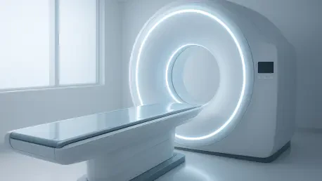

Total-body PET/CT represents a fundamental shift in medical physics, moving the industry away from the rigid trade-offs between image quality and scanning speed. For decades, the nuclear medicine community has been constrained by the physical limitations of short-axial field-of-view (SA-FOV) scanners, which typically cover only fifteen to twenty-five centimeters of the patient’s body at any given moment. This technological bottleneck meant that a full oncological assessment required the scanner bed to move through multiple positions, a process that could take up to twenty minutes and inherently wasted a significant portion of the radioactive signal emitted by the patient. The emergence of long-axial field-of-view (LA-FOV) systems, specifically the next-generation 128 cm total-body PET/CT scanners, has effectively shattered these constraints. By capturing the entire torso simultaneously, these systems harness a massive sensitivity dividend, allowing for the reconstruction of clear, diagnostic-grade images even when scanning times are reduced to a mere sixty seconds. This shift is currently redefining clinical workflows in 2026, offering a path toward higher throughput and significantly improved patient experiences without compromising the accuracy required for complex cancer care.
Methodological Rigor and Comparative Testing
Evaluating Performance: The Technical Scope of the EQUIP Study
The clinical evidence supporting this transition is best exemplified by the EQUIP study, which utilized a robust methodology to benchmark the performance of the GE HealthCare Omni 128 cm system. Researchers enrolled a cohort of twenty-four participants scheduled for standard-of-care scans using five distinct radiotracers, including Fluorine-18 FDG and Gallium-68 PSMA. This diversity was crucial for ensuring that the results were applicable across a broad spectrum of oncological applications, from metabolic glucose mapping to specific prostate cancer targeting. Each participant first underwent a standard scan on a conventional SA-FOV device to establish a baseline. They were then scanned for ten minutes on the LA-FOV system, but the real innovation lay in the data reconstruction process. The researchers meticulously broke down the total-body data into subsets that simulated five, three, two, and one-minute acquisition windows. This provided a granular view of how image quality persists even as the scanning time is radically compressed, allowing the team to identify the exact point where sensitivity compensates for the lack of duration.
Specialist Analysis: Blinded Reviews and Quality Scoring
To ensure that the results were not merely a product of technical optimism, the EQUIP study employed a rigorous blinded review process involving veteran nuclear medicine specialists. These experts were tasked with grading the reconstructed images on a five-point scale, focusing on diagnostic quality, image sharpness, and the presence of statistical noise. The results were remarkably consistent across all participants and tracers, providing a clear indication of the system’s superiority. A standout finding revealed that the one-minute reconstructions from the total-body system actually outperformed the full-length scans from conventional SA-FOV equipment. While the older technology earned respectable average scores, the ultra-fast total-body scans achieved higher marks for lesion conspicuity and overall clarity. This confirmed that the massive increase in detector coverage more than compensates for the eighty percent reduction in scanning time. By providing a perfect score for longer LA-FOV acquisitions and maintaining high scores for the sixty-second versions, the study proved that the “quality ceiling” for PET imaging has been raised to a level previously thought impossible.
Clinical Implications of Enhanced Sensitivity
Patient Comfort: The Critical Impact of Motion Management
One of the most immediate clinical advantages of ultra-fast total-body scanning is the radical improvement in motion management and patient comfort. In traditional PET imaging, the twenty-minute duration makes the acquisition highly susceptible to blurring caused by breathing, heartbeats, or the subtle shifting of a patient who may be in significant physical pain. By capturing a “metabolic snapshot” in just sixty seconds, the likelihood of such motion artifacts is effectively neutralized, leading to much sharper anatomical and functional alignment. This advancement is particularly transformative for vulnerable populations, including pediatric patients who often require sedation to remain still for traditional scans. Similarly, elderly patients or those suffering from claustrophobia or cognitive impairments find the one-minute scan much more tolerable, reducing the psychological burden associated with cancer staging. The ability to complete a diagnostic-quality scan within the time it takes to perform a simple breath-hold ensures that the clinical data remains consistent and high-fidelity, regardless of the patient’s ability to remain perfectly stationary for extended periods.
System Efficiency: Throughput and Radiation Safety
From an operational perspective, the sensitivity of the 128 cm LA-FOV system offers healthcare providers a powerful tool for optimizing both throughput and safety. In many metropolitan regions, access to PET/CT is a significant bottleneck in oncology, with waiting lists extending for weeks while patients wait for a slot on a conventional scanner. Reducing the time each patient spends on the table to just one minute allows imaging departments to potentially triple their daily capacity, clearing backlogs and accelerating the transition from diagnosis to treatment. Furthermore, this high sensitivity allows clinicians to make a different kind of trade-off: radiation dose reduction. Instead of using the sensitivity for speed, they can choose to inject a significantly lower amount of radiotracer while still maintaining standard image quality. This is a vital consideration for pediatric cases and for patients who require frequent follow-up scans over several years, as it minimizes the cumulative radiation exposure. The flexibility to prioritize either speed or safety depending on the clinical context makes total-body PET a versatile asset in modern health system management.
Future Directions and Technical Considerations
Accuracy Thresholds: Balancing Speed With Lesion Detection
While the initial data from the EQUIP study is overwhelmingly positive, the medical physics community in 2026 continues to maintain scientific caution regarding the limits of ultra-fast scanning. There is a meaningful distinction between “perceived quality,” where an image looks clear to a radiologist, and “diagnostic accuracy,” which involves the detection of microscopic or extremely faint lesions. Current research is focused on determining whether a one-minute scan might overlook very small metastases that require a higher count density to become visible against the background noise. Ensuring that speed does not come at the cost of sensitivity for early-stage disease remains a priority, and larger multi-center trials are currently underway to validate these findings across a wider range of pathology. The consensus is that while a one-minute scan is more than sufficient for general staging, certain complex cases may still benefit from longer acquisitions to ensure every potential site of disease is accounted for. This tiered approach to scan duration will likely become the standard, where the protocol is tailored specifically to the complexity of the patient’s condition.
Operational Logistics: Redesigning the Clinical Workflow
The successful integration of one-minute PET scans also highlights several logistical challenges that extend beyond the scanner room itself. Even if the PET portion of the exam is completed in sixty seconds, the patient still requires the standard hour-long “uptake time” to allow the radiotracer to distribute through the body. This creates a disconnect where the scanning hardware is much faster than the biological and administrative processes surrounding it. To truly capitalize on the speed of total-body PET, hospitals must redesign their clinic layouts, potentially increasing the number of uptake rooms and streamlining the check-in process to handle the increased patient flow. Furthermore, the massive amount of data generated by 128 cm scanners—often exceeding several gigabytes per patient—requires a significant investment in IT infrastructure and high-speed processing to prevent reconstruction times from becoming the new bottleneck. Moving forward, the broad adoption of this technology will depend as much on operational innovation and data management as it does on the underlying physics of the detectors.
The Path Toward Standardized Implementation
The EQUIP study firmly established that the transition from sectional to total-body imaging was a defining milestone in the evolution of nuclear medicine. By demonstrating that a sixty-second acquisition could exceed the quality of traditional ten-minute protocols, the researchers provided a compelling case for the widespread displacement of legacy hardware. This shift allowed for a fundamental reimagining of the oncology patient experience, where the stress of a long, confined procedure was replaced by a quick and efficient metabolic snapshot. The research also proved that the sensitivity of the 128 cm detector could be managed flexibly, offering a choice between increased throughput, reduced radiation exposure, or unmatched image clarity. As these systems became more common in major medical centers throughout 2026, the focus shifted toward optimizing their role within the broader cancer care pathway. The ability to capture the entire metabolic state of a patient in a single moment has set a new benchmark for clinical excellence, ensuring that the future of diagnostic imaging was defined by both unprecedented speed and surgical precision.