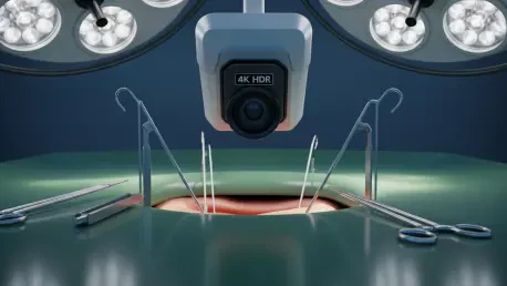

Surgeons operating within the high-intensity environments of modern operating rooms frequently encounter a frustrating visual paradox where high-output xenon lighting creates brilliant reflections on wet tissue while leaving adjacent anatomical recesses in total darkness. To address these critical visibility gaps, Vadzo Imaging has officially introduced the Falcon-821CRS, a high-performance 8MP (4K) surgical camera module designed specifically for the rigorous demands of healthcare visualization. This specialized tool, launched in Fort Worth, Texas, serves as a significant addition to the clinical imaging portfolio, targeting complex fields such as orthopedics, arthroscopy, and intraoperative procedures. By centering the design on high-end sensor technology and flexible connectivity, the development team has created a solution that prioritizes clarity and reliability in the most challenging lighting conditions. The launch represents a strategic effort to provide medical device manufacturers with a production-ready foundation that balances sophisticated image processing with a compact form factor. This release comes at a time when the demand for high-fidelity visual data is rising, driven by the increasing complexity of minimally invasive surgeries and the need for better tissue discrimination. Through the integration of the latest sensor innovations, the module ensures that both highlights and shadows retain their visual integrity, which is vital for maintaining surgical precision and ensuring patient safety during delicate maneuvers.
Illuminating the Void: Addressing Contrast in Surgical Environments
The physical environment of a modern surgical suite is characterized by extreme lighting contrast that frequently exceeds the technical capabilities of standard imaging sensors. High-intensity xenon and LED surgical lights are essential for providing deep cavity visibility, yet they often result in a “white-out” effect when they hit reflective surfaces like articular cartilage, metallic implants, or fluid-filled spaces. This glare can completely obscure the fine details of the anatomy, forcing surgeons to manually adjust lighting or reposition cameras, which can disrupt the flow of a procedure. In contrast, the darker recessed areas of a joint or cavity may remain underexposed, making it difficult to identify subtle pathologies or navigate small anatomical structures. The Falcon-821CRS was engineered specifically to mitigate these issues by providing a wide dynamic range that handles these lighting extremes simultaneously. By ensuring that the brightest highlights are not clipped and the deepest shadows remain visible, the camera allows for a more consistent and predictable visual experience for the surgical team.
Maintaining visibility during rapid lighting changes is a primary engineering hurdle that this new module effectively overcomes through sophisticated hardware-level processing. Rather than relying on post-processing software that can introduce significant lag or visual artifacts, the camera manages exposure levels in real-time. This is particularly important in arthroscopy, where the presence of irrigation fluids can create unpredictable reflections as instruments move within the joint space. The ability to maintain a clear view without flickering or brightness shifts allows the surgeon to focus entirely on the clinical task at hand rather than fighting with the imaging equipment. Moreover, this stability in image quality is essential for the accurate identification of tissue boundaries, which is a critical factor in successful orthopedic reconstructions. By providing a balanced image that replicates the natural perception of the human eye, the module enhances the overall confidence of the operating staff and reduces the cognitive load associated with interpreting degraded video streams.
Precision Engineering: The Capabilities of the HyperLux Sensor
At the technical core of this new imaging module is the Onsemi AR0821 HyperLux sensor, a 1/1.7-inch color CMOS component that sets a high bar for spatial resolution and sensitivity. With a native resolution of 3840 by 2160 pixels, the sensor provides the 4K clarity necessary for surgeons to distinguish between fine ligament fibers and subtle variations in tissue texture. The 2.1 micrometer pixel size is carefully calibrated to balance light gathering capability with high-resolution detail, ensuring that the system performs exceptionally well even when light levels are intentionally lowered to reduce heat generation at the surgical site. This sensor choice reflects a commitment to providing high-fidelity visual data that meets the standards of modern medical displays. The large sensor format also contributes to a better signal-to-noise ratio, which results in cleaner images with less graininess in darker areas of the frame. This clarity is not merely an aesthetic preference but a functional requirement for high-precision surgical navigation and instrument tracking.
One of the most critical features of the AR0821 sensor is its on-chip Line-Interleaved High Dynamic Range (LI-HDR) processing capability. Unlike traditional HDR methods that capture multiple frames and merge them through software, LI-HDR processes different exposure lines within the same frame at the pixel array level. This hardware-based approach is superior in medical environments because it eliminates the “ghosting” artifacts and motion blur that can occur when a surgeon moves an instrument quickly across the field of view. By capturing high-contrast data in a single, seamless stream, the Falcon-821CRS provides a more accurate representation of the surgical site than systems relying on conventional tone mapping. Furthermore, the integrated Image Signal Processor (ISP) automates critical functions such as auto-exposure, white balance, and noise reduction. This ensures that the raw data from the sensor is transformed into a clinically useful video stream without requiring extensive manual calibration by the hospital staff, thereby streamlining the setup process before each procedure.
Seamless Data Transmission: The Role of USB 3.0 Technology
To facilitate the transmission of high-bandwidth 4K video data without introducing latency, the Falcon-821CRS utilizes a robust USB 3.0 interface. This connection standard provides the necessary throughput to support 8MP resolution at frame rates that are high enough to ensure fluid motion on the surgical monitor. In the context of intraoperative visualization, any perceptible delay between the surgeon’s movement and the corresponding visual feedback can be dangerous and disorienting. By leveraging the high speed of USB 3.0, the module ensures that the visual loop remains tight, providing the real-time responsiveness required for delicate tasks like suturing or delicate tissue excision. The use of a standardized physical interface also means that the camera can be easily integrated into existing medical towers or portable diagnostic units without the need for specialized, expensive cabling. This move toward standardized high-speed connectivity reflects a broader industry trend toward interoperability and ease of hardware maintenance within the hospital environment.
Beyond raw speed, the module’s compliance with the USB Video Class (UVC) standard is a strategic advantage for medical device manufacturers and software engineers. This “plug-and-play” functionality allows the camera to be recognized as a standard video device by a wide range of operating systems, including Windows, Linux, and Android. For manufacturers, this eliminates the need to develop, maintain, and validate proprietary drivers, which can be a significant drain on research and development resources. In the medical field, where software updates must undergo rigorous testing to ensure they do not interfere with patient safety, the use of a standardized driver architecture significantly reduces the time and cost associated with regulatory qualification. This approach also ensures that the imaging system remains compatible with future operating system updates, protecting the hospital’s investment and reducing the risk of technical obsolescence. By simplifying the software layer, Vadzo Imaging allows medical device companies to focus their expertise on clinical applications and user interface design rather than basic hardware communication.
Clinical Superiority: Enhancing Outcomes in Specialized Procedures
The implementation of 4K HDR technology in the Falcon-821CRS offers tangible benefits across a variety of specialized surgical disciplines, particularly those that rely heavily on endoscopic visualization. In orthopedic and arthroscopic procedures, for instance, the ability to see the fine architecture of cartilage and the subtle nuances of ligament health is paramount for accurate diagnosis and repair. The high resolution allows for a greater degree of digital zoom without losing critical detail, enabling the surgeon to inspect small tears or degenerative changes that might be missed on a standard-definition system. Furthermore, the HDR capabilities prevent the aforementioned lighting “white-outs” when working near shiny surgical screws or instruments, ensuring that the surgeon never loses sight of the surrounding soft tissue. This consistent visibility is essential for ensuring that every maneuver is performed with the highest possible degree of accuracy, which is directly correlated with better long-term patient outcomes and faster recovery times.
Beyond traditional manual surgery, the high-fidelity output of this camera module is a vital asset for the growing field of robotic-assisted surgery and computer-aided navigation. These systems rely on stable, high-resolution visual sensors to track surgical instruments and map them against preoperative imaging data like CT or MRI scans. The clarity provided by the Falcon-821CRS ensures that the tracking algorithms can identify landmarks with a high degree of spatial accuracy, reducing the margin of error for robotic arms. Additionally, the lack of motion artifacts provided by the LI-HDR technology is crucial for AI-driven analysis tools that identify anatomical structures in real-time. If the video stream is blurry or has inconsistent lighting, the software may fail to correctly identify a critical nerve or blood vessel. By providing a clean and reliable data source, the module acts as the “eyes” of these advanced systems, enabling a higher level of automation and safety than was previously possible with older sensor technologies.
Structural Versatility: Designing for Medical Device Manufacturers
The physical architecture of the Falcon-821CRS is designed with a deep understanding of the spatial constraints inherent in medical device design. The module features a compact form factor that allows it to be integrated into a wide variety of enclosures, from overhead surgical lights to handheld diagnostic cameras and arthroscopic wand handles. Central to this versatility is the inclusion of an S-Mount (M12) lens holder, which is a standard in the embedded vision industry. This mounting system allows manufacturers to choose from a vast ecosystem of off-the-shelf and custom lenses, giving them the flexibility to tailor the camera’s field of view and focal depth to their specific clinical needs. For example, the same camera core could be used in a wide-angle overhead camera for general room visualization or a narrow-field, high-magnification lens for specialized dental or ophthalmic imaging. This modularity simplifies the supply chain for Original Equipment Manufacturers (OEMs), as they can qualify a single imaging platform for multiple product lines.
In addition to the hardware itself, Vadzo Imaging provides specialized engineering support to assist manufacturers throughout the integration process. This partnership is essential for addressing the unique challenges of the medical environment, such as the need for custom focus distances or specific mechanical adaptations to fit into proprietary surgical tools. Engineering teams can collaborate to optimize lens selection, ensuring that the optical path is perfectly matched to the sensor’s capabilities for maximum image sharpness. This support extends to the fine-tuning of the Image Signal Processor settings, allowing manufacturers to create a “signature look” for their video output that aligns with their clinical philosophy. By offering an end-to-end partnership, the company ensures that the camera module is not just a component, but a fully optimized imaging solution that meets the specific rigors of the healthcare sector. This collaborative approach reduces development risk and ensures that the final medical device meets the high standards required for clinical use.
Educational Advancement: Recording High-Fidelity Clinical Procedures
The high-grade 4K HDR video generated by the Falcon-821CRS plays a significant role in the advancement of medical education and professional development. In teaching hospitals, recording complex surgeries for later review is a standard practice, but the quality of these recordings has often been limited by the capabilities of the primary imaging sensor. By capturing every detail of a procedure in 4K resolution, the Falcon-821CRS allows students and residents to observe the nuances of tissue handling and instrument placement as if they were standing directly at the surgical table. The HDR aspect is particularly useful for education because it preserves the details of deep anatomical pockets that would normally be lost to shadow in a standard recording. These high-fidelity videos serve as an invaluable resource for creating training libraries, allowing the next generation of surgeons to study rare cases and perfect their techniques using the most accurate visual data available.
Moreover, the camera module facilitates the rise of remote proctoring and real-time surgical consultation, where experts can provide guidance from different geographic locations. The ability to stream 4K video through high-bandwidth hospital networks means that a consulting surgeon can see the same level of detail as the primary surgeon, ensuring that their advice is based on a clear and accurate understanding of the anatomical situation. This connectivity is further supported by the module’s ability to output at multiple resolutions, from 4K down to 1080p or even lower for mobile devices and tablets. This flexibility ensures that the video feed remains accessible even in environments with limited bandwidth, such as remote clinics or during mobile medical missions. By making high-quality surgical visualization more accessible and portable, the technology helps to democratize advanced surgical expertise and improve the standard of care on a global scale.
Strategic Implementation: Practical Steps for Clinical Adoption
Successful adoption of high-resolution imaging technology required a strategic approach that prioritized infrastructure compatibility and clinical validation. Institutions that transitioned to the Falcon-821CRS initially conducted comprehensive assessments of their existing surgical towers and display monitors to ensure they could support 4K HDR throughput. This proactive evaluation prevented bottlenecks in the data pipeline and ensured that the superior clarity of the sensor was fully realized at the point of care. Furthermore, hospital biomedical teams utilized the UVC compliance of the module to streamline the integration process, avoiding the common pitfalls associated with custom driver installations. These teams prioritized hardware that followed standardized protocols, which simplified the long-term maintenance of the equipment and reduced the frequency of software-related downtime. This focus on standardization proved to be a critical factor in maintaining high levels of operational efficiency within the operating suite.
Engineering departments within medical device companies took specific steps to leverage the mechanical flexibility of the S-Mount system, allowing them to iterate on product designs without changing the core imaging sensor. They collaborated closely with imaging specialists to select optics that minimized chromatic aberration and maximized the 8MP resolution of the Onsemi sensor. By standardizing on a single imaging core across multiple product lines, these organizations realized significant savings in documentation and regulatory compliance costs. They also integrated the camera’s HDR features into their automated tracking software, which resulted in more robust performance for robotic systems in challenging lighting conditions. Ultimately, the industry moved toward a model where high-fidelity visual data was treated as a foundational element of the surgical workflow, ensuring that every decision made in the operating room was supported by the clearest possible evidence. These actionable steps paved the way for a new era of precision in medical imaging, where technical limitations no longer dictated clinical possibilities.