
Despite the initial two cases serving as outliers due to a learning curve, the operator achieved stable and rapid performance by the third robotic procedure, a significant finding that comes from a rigorous assessment of robotic systems in the field of interventional neuroradiology where the stakes for precision and clinician safety have never been higher. Digital subtraction angiography (DSA) remains the undisputed gold standard for identifying complex vascular conditions such as intracranial aneurysms and arterial stenosis, yet the manual execution of these procedures places medical professionals in a precarious position. Traditionally, interventionalists must stand in high-radiation zones for extended periods, shielded only by heavy lead aprons that lead to chronic orthopedic issues and potential long-term health complications like malignancies or cataracts. The introduction of the YDHB-NS01 robotic system addresses these fundamental concerns by providing a remote-controlled platform that allows the surgeon to perform vascular navigation from a shielded control booth. By leveraging motorized precision and centralized command structures, this technology aims to decouple the physician from the hazardous environment of the angiography suite. This transition not only protects the surgical team but also introduces a level of mechanical consistency that promises to redefine procedural efficiency in modern neurosurgery.
Clinical Investigations: Study Design and Patient Benchmarks
The formal evaluation of the YDHB-NS01 Version 2.0 was conducted through a prospective clinical investigation at Peking Union Medical College Hospital throughout 2025. This study enrolled a total of 50 patients who required diagnostic cerebral angiography, purposefully dividing them into two equal groups of 25 to facilitate a direct comparison between robotic-assisted and manual methods. To ensure the integrity of the data and eliminate variables related to individual surgical expertise, a single interventional neurosurgeon performed all 50 procedures. This controlled approach allowed researchers to focus specifically on the interaction between the operator and the robotic hardware, measuring how the transition from manual tactile feedback to remote digital control influenced procedural metrics. The investigation was designed to test the limits of current robotic capabilities in a real-world clinical setting, providing a baseline for the safety and efficacy of the next generation of endovascular tools.
Rigorous baseline comparisons were conducted to ensure that the patient cohorts were statistically equivalent, preventing any demographic bias from skewing the results. Data extracted from the hospital’s electronic health records confirmed that there were no significant differences between the robotic and manual groups in terms of age, gender distribution, or the prevalence of systemic comorbidities like hypertension and diabetes mellitus. All procedures followed the standardized Seldinger technique for arterial access, typically utilizing the femoral or radial artery, which ensured that the initial stages of every surgery remained consistent with established medical protocols. Because the pre-procedural preparations were identical across both cohorts, the study effectively isolated the impact of the robotic slave unit on the intraoperative phase. This foundation allowed the clinical team to confidently attribute differences in procedural speed or navigation accuracy to the innovative hardware rather than variations in patient anatomy or baseline health status.
System Architecture: Master-Slave Hardware and Capabilities
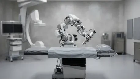
The technological foundation of the YDHB-NS01 system relies on a sophisticated master-slave architecture designed to translate human intent into robotic action with high fidelity. The system consists of a master console, where the surgeon is seated at a distance from the radiation source, and a bedside slave manipulator that directly engages with the patient’s vasculature. These components communicate via a secure local area network, ensuring that command inputs are transmitted with minimal latency and high-speed feedback is provided to the operator. The slave unit itself is a motorized assembly capable of manipulating standard, off-the-shelf diagnostic catheters, such as the five-French Cordis models used in the study. By utilizing existing clinical tools rather than proprietary catheters, the system lowers the barrier for hospital adoption and ensures that surgeons can continue to use the equipment they trust while benefiting from the added precision of robotic propulsion.
Several innovative features distinguish this robotic platform from earlier vascular assist devices, particularly in how it manages fluid dynamics and tool stability. The slave unit incorporates a dedicated guidewire holder that maintains perfect alignment during complex navigation, reducing the risk of accidental displacement that often occurs during manual manipulation. Furthermore, the system utilizes a unique one-way valve mechanism that facilitates continuous high-pressure infusion of heparinized saline or contrast agents. This design effectively eliminates the manual bottleneck of toggling three-way stopcocks, a task that traditionally requires a nursing assistant or a pause in the surgeon’s workflow. At the master console, the operator has centralized control over fluoroscopy activation, roadmap acquisition, and automated power injectors. This integration allows for a seamless execution of the angiography process, where imaging parameters and mechanical movements are managed from a single, ergonomically designed interface.
Operator Development: Simulation-Based Training for Proficiency
To prepare for the clinical trial, the participating neurosurgeon, categorized as a novice with fewer than three years of independent manual experience, underwent a comprehensive and structured training curriculum. This educational phase was critical for bridging the gap between traditional tactile surgery and the remote-control interface of the robotic system. The curriculum integrated theoretical modules on the electronic architecture of the YDHB-NS01 with hands-on practice using high-fidelity transparent vascular phantoms. These simulations allowed the operator to visualize the catheter’s behavior in a controlled environment, refining the subtle hand movements required to navigate the master handles without the pressure of a live clinical scenario. The training also emphasized the rapid setup of the sterile slave unit and the calibration of the motorized drive, ensuring that the transition from preoperative prep to active surgery was as efficient as possible.
Beyond basic navigation, the training protocol placed a heavy emphasis on patient safety and emergency preparedness. The operator was required to achieve mastery over manual override procedures, demonstrating the ability to immediately disconnect the robotic slave unit and take over the catheter manually if a mechanical failure or vascular complication occurred. Clinical application was only permitted after the surgeon demonstrated consistent technical proficiency across multiple simulated vascular anatomies. This focus on structured skill acquisition highlights a significant advantage of robotic technology: the potential to shorten the learning curve for junior clinicians. By providing a platform where movements are stabilized and imaging is centralized, the robot allows less experienced surgeons to achieve high standards of technical competence more rapidly than is often possible through manual apprenticeships alone.
Efficiency Metrics: Comparative Performance and Time Savings
The statistical results of the trial revealed that the robotic-assisted approach offered significant advantages in terms of procedural speed and workflow efficiency. While both the robotic and manual cohorts achieved a 100% technical and clinical success rate, the robotic group completed the active phase of the angiography significantly faster. The median procedure time—calculated from the moment the catheter entered the arterial sheath to its final withdrawal—was 27 minutes for the robotic cohort, compared to 38 minutes for the manual group. This 29% reduction in active surgical time suggests that the robotic interface allows for more direct and purposeful navigation of the cerebral vessels. Despite these gains, the total time patients spent in the angiography room remained comparable between the two groups. This was primarily due to the additional time currently required for the sterile draping and mechanical calibration of the robotic slave unit, which balanced out the time saved during the navigation phase.
The investigators identified three mechanical drivers behind the observed increase in procedural speed. First, the automated fluid delivery system significantly reduced the frequency of procedural pauses typically required for manual injections and saline flushes. Second, the stability provided by the robotic guidewire holder prevented the common manual frustration of losing a position within a tortuous vessel, which often necessitates repeated withdrawals and re-entries. Finally, the consolidation of all imaging and mechanical controls at the master console eliminated the “transit time” that manual operators spend moving between the patient’s bedside and the control monitors to review images. These features allowed the novice operator to maintain a continuous, uninterrupted workflow that outperformed the traditional manual method. Even when accounting for the radiation dose received by the patient and the volume of contrast used, the robotic system proved itself to be a highly efficient tool for diagnostic workflows.
Future Horizons: Addressing Limitations and Strategic Pathways
While the results were overwhelmingly positive, the clinical team recognized several areas where the technology must evolve to achieve universal adoption between 2026 and 2028. One of the primary limitations identified was the lack of sophisticated haptic feedback, as the current system does not yet transmit the subtle “feel” of the vessel wall to the operator’s hands. Additionally, the robotic system is currently restricted to the proximal and intermediate cerebral vasculature, with the navigation of distal microcatheters still requiring manual intervention in many cases. The initial arterial puncture also remains a manual task, indicating that the system is an assistant to the surgeon rather than a fully autonomous replacement. Future development cycles are expected to focus on integrating force-sensing technologies and expanding the system’s compatibility with a broader range of therapeutic microdevices to handle more complex interventions like clot retrieval or aneurysm coiling.
In the final analysis, the trial successfully established that the YDHB-NS01 system provides a safe and effective alternative to manual cerebral angiography. The complete absence of procedure-related complications, such as arterial dissections or thromboembolic events, reinforced the operational safety of the robotic platform. The rapid stabilization of the operator’s performance after only two cases demonstrated that the system is intuitive and accessible for the next generation of neurosurgeons. As the medical community moves toward broader implementation, the focus will likely shift toward multicenter trials and the development of standardized training certifications. The study concluded that the integration of robotic assistance significantly reduced the physical and radiological risks faced by medical staff while enhancing the speed of diagnostic procedures. These findings served as a clear indicator that the future of interventional neuroradiology will be defined by a synergy between human expertise and robotic precision, ultimately leading to safer environments for both physicians and patients.