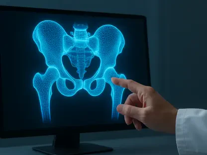

The transition from dense cortical bone to porous cancellous tissue presents a significant navigational challenge for surgeons performing delicate epiphyseal opening procedures. In the specialized field of pediatric orthopedics, this challenge is magnified by the high stakes of operating on a child’s skeletal system, where even a minor error can lead to lifelong consequences. The removal of physeal bars—bony bridges that cross the growth plate—requires a level of precision that traditionally relies on the surgeon’s subjective tactile feel and visual cues. However, as we look at the technological landscape of 2026, it is clear that the integration of artificial intelligence is redefining these boundaries. Current robotic systems have mastered trajectory planning and structural stability, yet they have historically lacked the nuanced sensory perception required to differentiate between varying bone densities in real-time. This deficiency creates a critical risk: over-grinding can destroy healthy growth tissue, while under-grinding leaves the clinical issue unresolved. By equipping surgical robots with a sophisticated autonomous recognition framework, researchers are now enabling these machines to perceive their environment with a granularity that surpasses human limitations. This transition from passive mechanical tools to active, sensing participants represents a fundamental shift in surgical methodology, ensuring that the next generation of pediatric procedures is governed by data-driven precision rather than variable human intuition.
The Physical Foundation: Multi-Source Sensing
To replicate the sophisticated touch of a veteran surgeon, robotic systems must go beyond simple visual monitoring and tap into the raw physical signals generated during the grinding process. When a high-speed surgical burr interacts with different bone structures, it produces a unique signature across multiple physical domains. This study utilized a tri-modal data stream, incorporating acoustic emissions, force dynamics, and micro-vibrations to create a high-fidelity representation of the tool-tissue interface. Acoustic signals provide immediate feedback on density changes, as the frequency and volume of the grinding sound shift the moment the burr exits the hard cortical shell and enters the softer cancellous bone. Simultaneously, force sensors track the mechanical resistance encountered by the tool, which drops significantly when the burr reaches more porous structures. By measuring these dynamics in conjunction with acceleration data that captures the tool’s vibrations, the system can build a comprehensive understanding of the material it is currently processing. This holistic approach ensures that the robot is not operating blindly based on a pre-loaded map, but is instead constantly updating its internal model based on the actual physical reality of the patient’s anatomy.
Building on this multi-layered sensory approach, the fusion of these distinct data streams addresses a primary weakness in single-sensor robotic designs. In a complex surgical environment, any single signal can be compromised by noise or mechanical interference; for example, a microphone might pick up ambient operating room sounds, or a force sensor might be influenced by the friction of the robot’s own joints. By integrating all three modalities—sound, force, and vibration—the framework creates a redundancy that ensures reliability even in less-than-ideal conditions. This multi-source information fusion allows the AI to cross-reference data points, validating a change in bone density across all three sensors before making a decision. This level of verification is essential for procedures like epiphyseal opening, where the margin for error is nearly non-existent. By synthesizing these inputs, the robot effectively gains a form of digital “tactile intuition” that remains consistent throughout the entire procedure, regardless of the surgeon’s fatigue level or the specific acoustic characteristics of the operating theater. This reliability is the cornerstone of the move toward more autonomous and responsive surgical platforms.
The Architecture: CNN-LSTM-Attention Model
The processing of such complex, simultaneous data streams requires a sophisticated computational architecture capable of handling both spatial and temporal patterns. The developed AI framework utilizes a three-channel parallel design where each sensory branch begins with Convolutional Neural Networks (CNN). These CNN layers are responsible for the immediate extraction of spatial features from the raw data, identifying specific patterns within the snapshots of information captured at every millisecond of the grinding process. However, bone grinding is not a static event; it is a continuous, time-dependent activity where the current state of the tissue is inextricably linked to what was encountered just moments before. To account for this, the model incorporates Long Short-Term Memory (LSTM) units, which allow the system to track and remember temporal features over the course of the operation. This ability to maintain a memory of the grinding trajectory is what enables the robot to pinpoint the exact moment of transition between bone layers with such high precision. Without this temporal awareness, a robot might misinterpret a brief spike in resistance as a change in bone type, leading to a premature or dangerous halt in the procedure.
A major innovation within this deep learning model is the “Squeeze-and-Excitation” attention mechanism, which serves as a dynamic prioritization system for the robot’s artificial senses. In a real-world surgical scenario, the relative importance of each sensor can change depending on the current task; the sound of the burr might be the most informative signal when it is spinning in open air, while the force sensor becomes the primary source of truth once the tool is deep within the cortical bone. The attention mechanism mimics human cognitive focus by dynamically re-weighting the importance of each sensory channel in real-time, effectively “listening” more closely to the sensor that provides the clearest data at any given moment. This adaptive weighting allows the network to filter out irrelevant noise and focus on the most critical information, ensuring that the recognition of the cancellous bone layer happens with maximum accuracy. By integrating spatial extraction, temporal memory, and adaptive attention, the model creates a robust decision-making framework that remains effective even as the physical conditions of the surgery fluctuate, providing a level of responsiveness that was previously impossible for robotic systems to achieve.
Achieving Precision: Real-Time Performance Metrics
The validation of this AI framework involved extensive testing with standardized artificial bone models, yielding results that set a new benchmark for surgical robotics. The CNN-LSTM-Attention model achieved a mean test accuracy of 95.06%, significantly outperforming traditional machine learning methods such as Random Forest or Support Vector Machines. To ensure that these results were not merely a product of memorizing specific datasets, the researchers employed a rigorous trial-by-trial data splitting method. This ensured that the model was tested on entirely separate grinding sessions that it had never encountered during its training phase, demonstrating its ability to generalize its knowledge to new and unique scenarios. The consistency of the performance was equally impressive, with a standard deviation of only 0.53% across independent runs, indicating that the system’s reliability is stable and predictable. This high level of accuracy is what provides the clinical confidence necessary to integrate such a system into pediatric surgery, where the robot must be able to distinguish between healthy and abnormal bone with near-perfect certainty.
In the fast-paced environment of the operating room, accuracy is only half of the equation; speed and latency are equally vital for patient safety. A surgical robot’s control system typically operates on a 50-millisecond loop, meaning that any AI-driven decision must be processed well within that timeframe to prevent the tool from moving too far into sensitive tissue. The proposed framework achieved an incredible inference time of just 1.2 milliseconds when running on an embedded platform like the NVIDIA Jetson Orin. This near-instantaneous processing speed means the robot can identify a tissue boundary and initiate a stop command or a motion adjustment almost the very moment the transition is detected. With only 0.32 million parameters, the model is lightweight and efficient, allowing it to be integrated into existing robotic hardware without requiring massive external computing power. This combination of extreme precision and low-latency performance ensures that the robot can act as a fail-safe against human error, immediately responding to the microscopic architecture of the bone faster than any human surgeon could ever react.
Clinical Implications: The Future of Orthopedics
The successful development and testing of this AI-driven sensing framework marked a significant milestone in the evolution of autonomous surgical assistants. By proving that a robot can accurately and instantly differentiate bone layers through sensor fusion, the research established a clear path toward safer and more effective pediatric orthopedic interventions. The study demonstrated that the combination of spatial and temporal data processing, enhanced by an attention mechanism, provides a level of environmental awareness that far exceeds the capabilities of traditional navigation-assisted robots. This transition from passive tools that follow a pre-set path to active participants that can “feel” their way through a procedure was validated as the new standard for high-precision surgery. The work highlighted the potential to significantly reduce the risk of growth plate damage in children, transforming a high-risk procedure into a more predictable and controlled medical event. The researchers also ensured that their findings were transparent and accessible, providing the broader scientific community with the datasets and models needed to further refine these technologies for diverse surgical applications.
Looking ahead, the focus must shift toward the seamless integration of these intelligent systems into live clinical workflows across various surgical disciplines. While the use of artificial bone models provided a robust proof of concept, the next step involves adapting the model to handle the complexities of living tissue, such as the presence of blood, irrigation fluids, and the natural anatomical variations found between individual patients. Hospitals and medical device manufacturers should consider this multi-source sensing framework as a foundational technology for a wide range of procedures, including spinal decompression and dental implant surgery, where tissue boundary recognition is equally critical. To fully realize the benefits of this technology, a collaborative effort is required to build larger, more diverse datasets that reflect a wider array of clinical scenarios. Implementing these AI models into the next generation of robotic platforms will not only enhance the safety of pediatric bone grinding but will also set a new global standard for surgical precision, where the fusion of human expertise and robotic sensing ensures the highest possible level of patient care. In the coming years, this approach will likely become the primary safeguard against the inherent risks of manual orthopedic procedures.