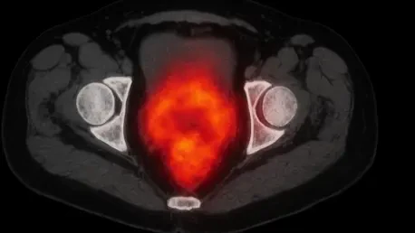

Gallium-68 PSMA-11 radiotracers create a vivid molecular signature that reveals metabolic activity even in anatomically normal regions of the prostate. This breakthrough addresses a persistent challenge in modern urology, where traditional multiparametric MRI often fails to detect high-grade tumors in roughly twenty to thirty percent of clinically significant cases. For many patients, the current diagnostic pathway becomes a source of extreme anxiety when rising prostate-specific antigen levels suggest disease, but imaging remains stubbornly inconclusive. This “diagnostic gray zone” frequently leads to a cycle of repeated and invasive systematic biopsies that sample the gland based on a generic grid rather than specific targets. Because these standard procedures are essentially blind to tumors hidden outside the typical sampling zones, aggressive cancers can go undetected while patients endure unnecessary risks of infection or bleeding. The latest research indicates that shifting the focus toward molecular imaging could finally close this dangerous gap for good.
Molecular Precision and Technological Innovation
The Biological Basis: PSMA Targeting
The FUPERMAN study fundamentally changed how clinicians view prostate diagnostics by shifting focus from anatomical distortions to the cellular behavior of the prostate-specific membrane antigen. This protein is heavily overexpressed on the surface of malignant cells, particularly in those that represent a high clinical risk. By utilizing the radiolabeled ligand gallium-68 PSMA-11, physicians can now pinpoint exact clusters of cancerous activity with unprecedented clarity. The intensity of the radiotracer accumulation often provides a direct window into the aggressiveness of the disease, as high-grade tumors generally demonstrate much higher PSMA levels than indolent tissues or healthy prostate cells. This molecular logic allows for the identification of “hot spots” that represent metabolic signatures of active cancer. Consequently, the technology bypasses the traditional reliance on physical tissue changes, offering a map based on biological function rather than just structural form.
Unlike multiparametric MRI, which relies on identifying physical distortions or changes in water diffusion within the tissue architecture, PSMA PET/CT illuminates the metabolic presence of the cancer itself. While MRI has long been the gold standard for its ability to identify lesions that distort the gland’s anatomy, its limitations are inherent in its medium; it cannot see what is not yet physically deformed. Molecular imaging, however, operates on a different plane by highlighting the chemical and biological markers of malignancy. This distinction is critical for early detection, as aggressive tumors may remain anatomically unremarkable during their most treatable stages. By visualizing the overexpression of PSMA, the medical community can now detect disease in regions that appear perfectly healthy on a standard scan. This evolution from structural to molecular observation represents a significant technological leap, ensuring that clinicians are no longer flying blind when an MRI returns a false negative result.
Implementing Fusion-Guided: Biopsy Techniques
To translate these molecular insights into clinical action, researchers implemented a fusion-guided biopsy approach that synchronized PET/CT data with real-time ultrasound. This methodological rigor was applied to a specific cohort of sixty-three men who shared a common diagnostic dilemmnegative or inconclusive MRI results despite a high clinical suspicion of cancer. During the procedure, the molecular “heat map” generated by the gallium-68 PSMA-11 tracer was digitally overlaid onto the live ultrasound feed used by the urologist. This co-registration allowed for a level of surgical precision previously unattainable in patients with clean MRIs. By having a clear target illuminated by the radiotracer, the medical team could ensure that biopsy needles were directed specifically at the most metabolically active sites. This approach effectively neutralized the guesswork associated with traditional sampling, transforming the biopsy from a probabilistic grid search into a targeted, data-driven surgical intervention.
The success of this technique relied heavily on the seamless integration of different imaging modalities within a single procedural workflow. By comparing this targeted method against the current standard of systematic biopsy in a controlled environment, the study provided a clear framework for evaluating the “yield” of molecular guidance. The dual approach ensured that every participant received both the targeted samples and the traditional grid-based cores, allowing for a direct head-to-head comparison of their effectiveness. This experimental design highlighted how real-time data fusion can overcome the limitations of anatomical imaging. Furthermore, a subset of patients participated in exploratory protocols using dynamic total-body PET, which tracked the kinetic flow of the radiotracer over time. This additional layer of temporal data suggested that the speed and duration of tracer binding could offer even deeper insights into tumor behavior, paving the way for a new era of highly sophisticated, precision-guided urological surgery.
Validating Superior Detection Rates and Future Standards
Breakthroughs: Cancer Identification
The results of the FUPERMAN study demonstrated that PSMA PET/CT-targeted biopsies were significantly more effective than conventional methods at identifying aggressive disease. Specifically, the molecularly guided approach alone uncovered clinically significant prostate cancer in thirty-seven percent of the study population, nearly doubling the success rate of the twenty-one percent achieved by systematic sampling. When clinicians combined both methods, the detection rate climbed to forty-one percent, proving that molecular imaging captures tumors that fall entirely outside the standard biopsy grid. These findings were particularly striking because the imaging showed a sensitivity of nearly ninety-six percent, identifying almost every patient who was eventually confirmed to have high-grade disease. By consistently illuminating what was previously invisible, the technology reduced the likelihood of false negatives and ensured that aggressive tumors were caught early enough for potentially curative treatment.
Beyond the binary success of detection, the research team focused on how quantitative metrics like the maximum standardized uptake value, or SUVmax, served as a reliable indicator of tumor severity. This measurement allowed clinicians to stratify patient risk with a high degree of confidence, as higher concentrations of the radiotracer were directly linked to more aggressive cellular profiles. The ability to distinguish between slow-growing, indolent tissues and dangerous, high-grade cancers meant that patients could be managed with greater personalization. For those with low molecular uptake, the results provided a clear rationale to avoid more invasive interventions, while those with intense “hot spots” were fast-tracked for surgery or radiation. This refinement of risk assessment represented a major step forward in modern oncology, moving away from generalized treatment protocols toward a model where every medical decision is informed by the specific molecular profile of the individual patient’s tumor.
Future Outlook: Implementation Challenges
The researchers concluded that while the initial findings were promising, the path to widespread adoption required navigating several practical and economic hurdles. It was determined that the high cost of PET/CT equipment and the specialized production of gallium-68 PSMA-11 radiotracers needed to be addressed to ensure equitable access across different healthcare systems. Hospitals were encouraged to begin investing in the multidisciplinary training required for radiologists, nuclear medicine specialists, and urologists to work in concert. Furthermore, the study suggested that future efforts should focus on large-scale, multi-institutional trials to confirm these detection rates across more diverse populations. By establishing a robust evidence base, the medical community sought to prove that the initial investment in molecular imaging would eventually lower long-term costs by reducing the number of repeated, failed biopsies and improving the overall success rate of early cancer interventions.
Looking ahead, the integration of molecular imaging into standard clinical guidelines was viewed as a necessary evolution for the management of the diagnostic gray zone. Practical next steps involved streamlining the fusion-guided workflow to make it more accessible for community-based urology practices rather than just academic centers. The healthcare industry recognized that providing a clear diagnostic alternative for men with negative MRIs could drastically reduce the emotional and physical burden of diagnostic uncertainty. Solutions were proposed to optimize the distribution of radiotracers and to refine the software used for real-time image co-registration. Ultimately, the study provided a blueprint for a more accurate and compassionate diagnostic pathway, where molecular maps lead clinicians directly to the disease. By prioritizing these technological and logistical advancements, the medical field moved closer to a future where aggressive prostate cancer is no longer hidden by the limitations of anatomical imaging.