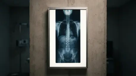

Researchers noted that plain radiographs often lack the sensitivity required to detect early-stage osteoporosis or cancer, which can lead to a dangerous sense of false reassurance. This critical insight underscores the ongoing tension between traditional chiropractic methods and modern, evidence-based medical standards. For decades, the chiropractic profession has utilized spinal imaging not only to screen for severe pathologies but also to analyze biomechanical alignment and determine specific vectors for manual adjustment. However, contemporary clinical guidelines across the healthcare spectrum have increasingly cautioned against routine imaging for nonspecific spinal pain, citing a lack of evidence that such procedures lead to improved patient outcomes. To address this persistent gap in clinical knowledge, a specialized research team led by Hazel J. Jenkins in Sydney, Australia, initiated a pilot trial designed to evaluate the feasibility of a full-scale randomized controlled trial. This effort was grounded in the principle of clinical equipoise, a state where the medical community remains genuinely uncertain about which diagnostic path offers the most substantial benefit to the patient. By investigating the logistical and practical hurdles of such a study, the researchers sought to establish a framework that could eventually determine if spinal radiographs truly enhance health and safety or merely add unnecessary layers to the care process.
Clinical Discrepancies: Navigating Policy and Professional Practice
The current landscape of chiropractic practice is characterized by a significant divergence between official recommendations and real-world application. Most national and international guidelines suggest a conservative approach, advising practitioners to reserve spinal radiographs for cases involving “red flags,” such as suspected fractures, malignancies, or systemic inflammatory conditions. Despite these clear directives, the actual frequency of imaging referrals in the field remains remarkably inconsistent. Data collected from various clinical settings indicates that referral rates can fluctuate wildly, with some practitioners ordering X-rays for only 8 percent of their new patients while others maintain a rate as high as 84 percent. This massive variance often reflects individual philosophical leanings rather than purely objective clinical needs, highlighting a professional culture that is still grappling with the transition toward a standardized, evidence-led model of care.
This inconsistency creates a complex dilemma for modern clinicians who must balance diagnostic thoroughness against the potential harms of overutilization. On one hand, the inappropriate use of radiographic imaging introduces several documented risks, including unnecessary exposure to ionizing radiation and the financial burden of procedures that may not alter the treatment plan. Furthermore, there is the persistent risk of overdiagnosis, where benign or age-related findings are misinterpreted as significant drivers of pain, leading to patient anxiety or unnecessary interventions. On the other hand, many chiropractors harbor a deep-seated fear that failing to perform a pretreatment scan might cause them to miss a subtle contraindication, potentially resulting in an adverse event during spinal manipulative therapy. This tension between radiation safety and diagnostic certainty remains at the heart of the debate, necessitating rigorous scientific inquiry to provide practitioners with the clarity needed to make informed decisions.
Methodology and Selection: Finding the Middle Ground
To ensure the integrity of the pilot study, the research team implemented a rigorous screening process to identify chiropractors who genuinely occupied a “zone of clinical equipoise.” This meant selecting practitioners who were neither strictly opposed to imaging nor reflexively dependent on it for every case. The inclusion criteria were limited to registered chiropractors in the Sydney region who maintained a moderate imaging referral rate—specifically between 20 percent and 80 percent of their new patients. This range was strategically chosen because clinicians with extreme referral patterns likely have entrenched views that would make them less likely to adhere to a strict randomization protocol. Out of 40 clinicians initially screened, only 16 met these specific criteria, and ultimately, 11 consented to participate in the trial. This relatively small recruitment pool highlighted the difficulty of finding practitioners who are comfortable navigating the uncertainty inherent in randomized diagnostic trials.
The participating chiropractors underwent a comprehensive training program designed to prepare them for the nuances of the study. This training focused on identifying diagnostic uncertainty and utilizing specific communication scripts to explain the concept of randomization to their patients. One of the primary goals was to ensure that discussing the “unknown” necessity of an X-ray did not undermine the therapeutic relationship or the patient’s trust in the clinician’s expertise. By standardizing the way uncertainty was framed, the researchers aimed to minimize bias and ensure that the decision to image was driven by the study protocol rather than the practitioner’s subconscious preferences. This methodological foundation was essential for testing whether a larger trial could successfully navigate the psychological and professional barriers that often prevent high-quality research in a private clinical setting.
Patient Safety: Protocols and Participant Profiles
The recruitment of patients focused on adults presenting with new episodes of spinal pain, defined as symptoms occurring in individuals who had not experienced similar issues in the preceding month. To maintain the highest safety standards, the study excluded anyone under the age of 18, pregnant women, or individuals displaying any “red flag” symptoms that would mandate immediate imaging. This careful selection ensured that the participants were individuals for whom the decision to image was truly a matter of clinical judgment rather than an emergency necessity. To bridge the gap between research and care, all eligible patients were provided with a sealed imaging referral before they were randomized. Those assigned to the imaging group were instructed to proceed with the scan immediately, while those in the control group were told to hold the referral and only use it if their clinical situation deteriorated or failed to show progress.
Despite the careful design, the study faced significant logistical challenges, primarily due to external disruptions that occurred during the recruitment phases between early 2026 and mid-2027. Out of a screened cohort of 135 patients, only 20 individuals ultimately completed the randomization process and baseline requirements. The demographic data of these participants revealed a relatively young population, with a mean age of 36, reporting moderate pain levels but low functional disability. Interestingly, more than half of these patients entered the study with a preexisting belief that radiographs were a mandatory component of high-quality spinal care. This cultural expectation suggests that the demand for imaging is often driven as much by patient perception as by clinical need, further complicating the practitioner’s role in implementing evidence-based guidelines that might suggest an X-ray is unnecessary.
Clinical Realities: Examining Crossover and Outcomes
A significant aspect of the pilot trial involved monitoring group compliance, specifically looking for “crossover” where participants did not follow their assigned diagnostic path. The study found that 30 percent of the participants in the imaging group chose not to obtain their X-rays because their symptoms improved so rapidly that the procedure no longer felt justified. Conversely, 20 percent of the non-imaging group ended up receiving scans within the first two weeks due to a lack of progress or a worsening of their condition. These instances of crossover illustrate the fluid and dynamic nature of clinical practice. They demonstrate that while a research protocol provides a roadmap, the evolving health status of the patient often necessitates a departure from the plan. This observation is crucial for designing future trials, as it highlights the need for flexible statistical models that can account for real-world clinical decision-making.
The health outcomes measured during the 12-week follow-up period were overwhelmingly positive for both the imaging and control groups. Participants across the board reported significant decreases in pain intensity and notable improvements in functional mobility. Furthermore, patient satisfaction remained exceptionally high, with 94 percent of the cohort expressing high or very high satisfaction with the care they received by the six-week mark. Safety data supported the conclusion that the absence of initial radiographs did not lead to increased risk, as adverse events were rare and limited to transient issues like muscle soreness or temporary reaggravation of pain. These findings suggested that for the majority of patients with nonspecific spinal pain, the timing of radiographic imaging did not appear to be a primary determinant of short-term recovery or treatment safety.
Strategic Evolution: Reframing Research for Professional Integrity
The qualitative phase of the study involved in-depth interviews with the participating chiropractors, which shed light on the psychological challenges of conducting diagnostic research. Many clinicians admitted that admitting to “clinical uncertainty” regarding the necessity of an X-ray felt like an erosion of their professional authority. In an environment where patients often seek definitive answers for their discomfort, the practitioners expressed a sense of vulnerability when they could not provide a clear justification for why a scan was or was not being performed. This feedback emphasized that for a full-scale trial to succeed, the responsibility for explaining randomization and equipoise should be shifted to dedicated research staff. This adjustment would allow the treating chiropractor to maintain their role as a confident provider of care while the research team handles the complexities of the study’s scientific requirements.
The pilot trial successfully identified the structural modifications required to transition into a definitive, multicenter investigation. The researchers established that while the safety and follow-up protocols were robust, future efforts must focus on diversifying the participant demographic to include older populations who may present with different clinical risks. The study also highlighted the necessity of addressing the ingrained societal beliefs about the utility of diagnostic technology through better patient education. By refining the recruitment strategies and removing the communicative burden from the clinicians, future research will be better positioned to provide a final answer on the value of spinal imaging. This progression represented a vital step toward reconciling traditional chiropractic practices with the demands of modern medicine, ensuring that diagnostic tools are used judiciously to maximize patient benefit while minimizing unnecessary risks.