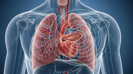

The rapid evolution of diagnostic imaging often outpaces the development of standardized communication protocols, leaving clinicians to navigate a complex landscape of varying terminology and interpretive nuances during life-threatening emergencies. Acute pulmonary embolism remains one of the most daunting challenges in the emergency department, where every second spent deciphering a radiology report can directly impact a patient’s survival probability. To address this critical friction point, the American College of Radiology has introduced the Pulmonary Embolism Reporting and Data Systems, or PE-RADS v2026, marking a significant transition toward structured, data-driven diagnostics. Published in the journal Radiology, this framework provides a universal blueprint for documenting pulmonary vascular obstructions, ensuring that the findings identified by imaging specialists are immediately understood and acted upon by the entire clinical team.
Bridging the Diagnostic Gap: Building a Shared Clinical Language
To appreciate the necessity of PE-RADS, it is vital to understand the clinical gravity of acute pulmonary embolism, a condition where a blood clot suddenly blocks a pulmonary artery and causes immense strain on the heart. Because the window for effective intervention is frequently narrow, the accuracy and speed of the initial diagnostic report are paramount to ensuring the patient receives the correct level of care. Historically, the absence of a unified reporting structure led to variations in how findings were prioritized, creating potential delays in treatment. To solve this, the American College of Radiology partnered with front-line clinicians from the PERT Consortium and the American College of Emergency Physicians to build a consensus. By establishing a rigorous foundation for reporting, the new system helps to minimize risks by focusing on the most critical clinical indicators that determine patient outcomes in real time.
Enhancing Communication: The Role of Terminology in Acute Care
The primary mission of the PE-RADS v2026 framework is to eliminate the ambiguity and inconsistency often found in traditional radiology reports through a highly structured and predictable reporting format. In the past, radiologists might use differing terminology to describe the severity or location of a clot, which could lead to confusion among the multidisciplinary teams—including emergency physicians and pulmonologists—who must act on those findings. By establishing a structured lexicon, PE-RADS provides a clear and consistent language that ensures every member of the care team understands the clinical implications of the imaging results. This shift toward standardization means that regardless of which facility a patient visits, the information conveyed to the attending physician will follow a logic that is universally recognized. This reliability is particularly important for junior residents who may be interpreting scans.
Streamlining Response: Integrating Reports into Hospital Workflows
Beyond just word choice, the standardization offered by PE-RADS v2026 accelerates the decision-making process by streamlining how critical findings are flagged within the electronic health record system. When a radiologist categorizes a case using the standardized descriptors, the emergency team can immediately recognize the severity of the obstruction and the potential for right ventricular dysfunction. This shared mental model reduces the need for lengthy follow-up phone calls and clarifications, allowing the medical team to focus on preparing for thrombolysis or surgical intervention. Moreover, the consistency in reporting enables the seamless transfer of information when patients move from the emergency department to the intensive care unit or the operating room. This continuity is essential for managing complex cases where multiple specialists are involved in the journey, ensuring the signal of an emergency is heard clearly.
Technical Framework and Risk Assessment: From Imaging to Action
Standardization requires a deep integration of both anatomical findings and physiological responses, which is exactly what the technical framework of PE-RADS achieves by linking clot location to heart health. The system utilizes a sophisticated approach to categorize findings based on their hemodynamic significance, ensuring that a simple clot is distinguished from a massive embolism causing immediate heart failure. This dual focus allows the imaging report to serve as a comprehensive risk assessment tool rather than a mere list of anatomical observations. By incorporating the latest research on right ventricular strain, the framework provides a more holistic view of the patient’s condition, which is vital for choosing between conservative management and invasive treatments. This shift marks a major milestone in cardiovascular diagnostics, where the radiologist’s role is increasingly intertwined with the management of acute vascular diseases.
Defining the Grading Scale: Classification and Stratification
At the heart of the PE-RADS system is a robust 0–4 grading scale that allows radiologists to classify the location of the clot while simultaneously integrating critical heart imaging features into a single score. This hierarchical framework facilitates risk stratification, the process of determining which patients are at the highest risk of cardiac collapse and require aggressive intervention versus those who can be managed with standard treatments. By evaluating the right heart’s response to the embolism, the system provides a clear roadmap for immediate clinical next steps based on objective data rather than subjective clinical intuition. Each level of the scale corresponds to a specific set of clinical recommendations, which helps to standardize the treatment protocols across different hospital systems. This prevents over-treatment of low-risk patients while ensuring those with life-threatening emboli receive the fastest possible care.
Managing Technical Constraints: Improving Image Reliability and Accuracy
Furthermore, the system accounts for the technical complexities of modern imaging by including specific modifiers for image quality issues, such as motion artifacts or poor contrast that might obscure a diagnosis. While CT pulmonary angiography remains the primary diagnostic tool, the framework also encompasses MR angiography to provide high-accuracy alternatives for specific patient needs, such as contrast allergies. By identifying non-PE causes of arterial abnormalities and providing guidance on technical limitations, PE-RADS ensures that the final diagnosis is as precise and actionable as possible. This transparency regarding the quality of the image allows clinicians to weigh the reliability of the report against the patient’s clinical presentation, fostering a more nuanced approach. If an image is suboptimal, the system provides directions on whether additional imaging or alternative diagnostic pathways should be pursued to avoid dangerous delays.
Advancing Clinical Outcomes: Standardized Protocols for Long-Term Safety
The implementation of the PE-RADS v2026 system represented a decisive shift toward clinical precision and interdisciplinary transparency in the management of one of medicine’s most acute emergencies. Healthcare institutions that integrated these standards witnessed an immediate improvement in the speed of communication between radiology departments and emergency units, which directly translated to faster treatment times for critical patients. Moving forward, clinical leaders were encouraged to incorporate these standardized templates into their existing electronic health record systems to maximize the efficiency of the new protocol. The focus shifted toward training all medical staff on the nuances of the grading scale, ensuring that the benefits of the framework reached every level of patient care. By adopting this structured approach, providers not only enhanced the safety of current diagnostic workflows but also contributed to a global database that refined treatment pathways.