
The ability to monitor the wash-in and wash-out phases of a contrast agent in real time is essential for navigating the complicated diagnostic landscape of liver disease. This environment is frequently clouded by the presence of “imposter” lesions—masses that mimic the appearance of malignancy on standard imaging but remain fundamentally benign. Among these, solitary necrotic nodules, or SNNs, present a unique challenge to clinicians because they consist of dead tissue that can easily be mistaken for metastatic disease or hepatocellular carcinoma. Researchers at the First Hospital of Jilin University have recently conducted a landmark study to demystify these lesions. By analyzing eighty-seven necrotic liver lesions across eighty-one patients, the team has successfully identified specific characteristics that set these nodules apart. This breakthrough is transformative for hepatology, as it provides a non-invasive pathway to clear diagnostic certainty and reduces the psychological toll on patients during routine screening.
Diagnostic Innovations and Advanced Imaging
Enhancing Precision: The Role of Microbubble Technology
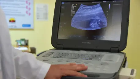
Contrast-enhanced ultrasound, or CEUS, represents a fundamental shift in how physicians evaluate liver masses at the bedside. The technology utilizes an intravenous injection of microscopic, gas-filled bubbles that act as purely intravascular tracers. Unlike traditional contrast agents used in other imaging modalities, these microbubbles do not leak into the interstitial space, allowing for an incredibly precise visualization of the organ’s vascular architecture. This is particularly relevant for the liver, which possesses a dual blood supply from both the hepatic artery and the portal vein. The real-time nature of CEUS enables radiologists to observe how contrast moves through a lesion second by second. For solitary necrotic nodules, which are essentially islands of dead tissue, the absence of internal blood flow becomes immediately apparent. This lack of vascularity is a primary indicator of a benign state, contrasting with the aggressive blood vessel networks found in malignant tumors.
Beyond its diagnostic precision, CEUS offers several practical advantages over more conventional imaging techniques such as computed tomography or magnetic resonance imaging. One of the most significant benefits is the complete absence of ionizing radiation, making it a safer option for repeated follow-up examinations or for patients with sensitivities to radiation exposure. Furthermore, the procedure can be performed rapidly in various clinical settings, including outpatient clinics or even at the bedside for immobilized patients. While MRI provides excellent detail, it often requires lengthy scan times and involves claustrophobic environments that many patients find distressing. In contrast, CEUS provides immediate functional data without the need for extensive preparation or long waiting periods. This accessibility, combined with the safety profile of the microbubbles, ensures that patients can receive a definitive assessment of suspicious nodules without the risks of nephrotoxicity.
Recognizing Morphological Signatures: Shape and Structure
A critical finding from the Jilin University study involves the specific physical geometry of solitary necrotic nodules compared to other types of liver masses. While malignant tumors often present as relatively neat, spherical, or oval lumps, SNNs frequently exhibit irregular and complex shapes. The research demonstrated that seventy-seven percent of the analyzed lesions were irregular, with a significant majority displaying a tortuous, cord-like configuration. This appearance suggests a history of localized tissue death that follows the path of a specific vessel or duct rather than the radial growth characteristic of a tumor. Recognizing these “twisted” or “stringy” shapes on a scan allows a radiologist to move away from a cancer diagnosis early in the evaluation process. These morphological cues serve as the first line of defense against diagnostic errors, providing a structural signature that points toward a benign, necrotic origin rather than a proliferative malignancy.
In addition to external shape, internal markers visible on standard grey-scale ultrasound provide further diagnostic clues for identifying solitary necrotic nodules. The research team identified central hyperechoic foci—bright, reflective spots located in the middle of the mass—in a notable portion of the studied cases. These bright spots often arrange themselves in “equal-sign-like” or “core-type” patterns, which are rarely seen in aggressive tumors. Such reflections are thought to represent residual fibrous tissue or tiny calcifications within the necrotic core. When these internal markers are paired with the irregular, cord-like shapes previously mentioned, the probability of the lesion being a benign SNN increases substantially. This detailed cataloging of internal and external features creates a more robust diagnostic framework. It allows clinicians to look beyond the mere presence of a mass and instead focus on the specific architectural nuances that define the difference between a harmless patch of dead tissue and a growth.
Analyzing Contrast Patterns and Clinical Care
The Significance of the Peripheral Rim: Border Characteristics
When the contrast agent is introduced during a CEUS examination, the behavior of the lesion’s border becomes a primary focus for diagnostic differentiation. The study revealed that nearly forty-five percent of solitary necrotic nodules exhibit what is known as a “rim of hyperenhancement” during the arterial phase. This phenomenon occurs when the outer edge of the nodule lights up on the screen while the central portion remains dark and unenhanced. This bright rim represents the living tissue—often a mix of inflammatory cells or fibrous material—that the body naturally builds as a wall around the dead necrotic core. It is a biological response to the presence of dead tissue, essentially an attempt to sequester the area from the rest of the healthy liver. Understanding that this enhancement is confined to the periphery is vital, as it confirms that the interior of the mass is truly avascular. This specific pattern is a hallmark of necrosis and serves as a strong indicator that the lesion is not a solid tumor.
The thickness of this peripheral rim serves as a critical variable that can complicate or clarify the diagnosis. The researchers found that while many rims are thin and barely perceptible, roughly twenty-eight percent of the cases featured “thick rims” measuring five millimeters or more. This thickness is statistically significant because it changes the way the nodule interacts with the contrast agent as the scan progresses into the portal venous phase. Thick rims are often associated with more intense inflammatory activity or extensive fibrosis at the lesion’s border. Interestingly, when contrast is applied, these nodules can sometimes appear to “grow” in size because the enhancing border defines a larger visible boundary than the initial grey-scale scan suggested. By establishing a five-millimeter threshold for “thick” rims, the study provides clinicians with a quantifiable metric to assess the lesion. This measurement helps predict how the nodule will behave in subsequent phases of the imaging process.
Distinguishing Benign Washout: Improving Clinical Outcomes
One of the most treacherous aspects of liver imaging is the phenomenon known as “washout,” where a lesion loses its contrast faster than the surrounding healthy liver tissue. In traditional radiology, washout is considered a primary indicator of malignancy, particularly for hepatocellular carcinoma. However, the study discovered that this rule is not absolute and can be misleading when evaluating necrotic nodules. Specifically, the research showed that nearly half of the solitary necrotic nodules with thick rims also exhibited washout during the portal venous phase. This mimicking of cancer behavior occurs because the fibrous or inflammatory border loses contrast more rapidly than the liver’s normal blood-rich tissue. Without the context provided by this study, a radiologist might see this washout and incorrectly conclude that the patient has a malignant tumor. This finding is a major breakthrough because it warns medical professionals that a benign lesion can look like cancer if its surrounding border is sufficiently thick.
Moving forward, the medical community successfully integrated these ultrasound techniques into standard diagnostic protocols to enhance accuracy in hepatology. Clinicians prioritized the identification of morphological markers, such as the cord-like configuration and internal hyperechoic foci, as primary indicators of benign solitary necrotic nodules. They also adopted the five-millimeter rim thickness threshold to better interpret deceptive washout patterns, which significantly decreased the rate of false-positive cancer diagnoses. This progress encouraged a broader implementation of contrast-enhanced ultrasound across diverse healthcare settings, ensuring that patients benefited from real-time vascular analysis without the need for radiation-heavy scans. By focusing on these specific diagnostic signatures, healthcare providers effectively reduced the necessity for invasive procedures while maintaining a high level of vigilance. Ultimately, these advancements established a more efficient framework for managing liver findings, ensuring that the focus remained on patient health.