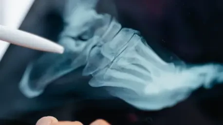

The diagnostic accuracy for identifying medium- to high-risk complications in heel surgery reached only 71.5 percent when radiologists worked without technological assistance. The calcaneus, or heel bone, serves as the primary weight-bearing structure of the foot, often shattering into a complex labyrinth of fragments during high-energy trauma. To repair such fractures, surgeons must employ an intricate arrangement of metal plates and screws, which subsequently creates a dense, overlapping visual field on standard postoperative X-rays. Identifying subtle complications like joint penetration or hardware instability within this chaotic 2D landscape is a notorious challenge for radiologists. Recent research led by Shaobin Wang and Hai Li has introduced GPT-5 as a sophisticated diagnostic co-pilot to address this clinical bottleneck. By integrating multimodal large language models into the radiology workflow, this study sought to determine if AI could provide the necessary safety net to improve diagnostic reliability while reducing the heavy cognitive load placed on specialists who must navigate these high-stakes orthopedic cases.
Enhancing Diagnostic Accuracy through AI Integration
Innovative Study Methodology: A Multicenter Approach
To assess the impact of this technology, the research team conducted a multicenter, multi-reader study involving six radiologists with varying levels of professional experience. These experts analyzed 73 specific cases of postoperative calcaneal fixation, resulting in 438 individual observations. To establish a ground truth, each interpretation was compared against a pragmatic reference standard, which involved postoperative CT scans reviewed by a consensus of expert radiologists. This design ensured that the findings were grounded in clinical reality, as CT remains the gold standard for identifying hardware issues that might be obscured on traditional X-rays. The study was structured into two distinct phases: an initial session where doctors worked entirely unaided, and a subsequent session following a four-week washout period. During the second phase, the participants were provided with draft reports and structured checklists pre-generated by GPT-5, allowing them to review and refine AI-suggested findings rather than starting from a blank page.
Significant Gains: Improving Sensitivity and Detection Rates
The performance data gathered during the assisted session demonstrated a significant leap in the ability to identify complications requiring surgical escalation. While the baseline accuracy of 71.5 percent without AI was representative of current clinical challenges, the integration of GPT-5 boosted this figure to 80.1 percent. Statistically, this shift nearly doubled the odds of a radiologist’s decision aligning with the expert CT consensus, highlighting a major reduction in diagnostic uncertainty. Most notably, the sensitivity of the readers—their capacity to correctly flag a genuine problem—surged from 54.2 percent to 79.2 percent. This suggests that the AI draft reports acted as an effective visual guardrail, pointing out subtle abnormalities that might be overlooked in the dense overlay of metal and bone. By alerting the doctor to potential misses, the system effectively lowered the threshold for detecting medium- to high-risk issues that would otherwise necessitate a more expensive and invasive diagnostic path for the patient.
Optimizing Workflow and Radiologist Confidence
Drastic Efficiency: Reducing Reading Times in Clinical Settings
One of the most immediate benefits observed in the study was the dramatic acceleration of the reporting process. In the unaided session, radiologists required an average of 378.6 seconds—over six minutes—to interpret a single case and document their findings. With the assistance of GPT-5, this time plummeted to just 98.4 seconds per case. This nearly fourfold increase in speed has profound implications for modern healthcare environments, where high patient volumes and radiologist shortages are driving widespread professional burnout. By providing a pre-populated draft report, the AI handles the repetitive task of formalizing descriptions, allowing the clinician to shift their focus toward verifying specific anatomical landmarks. This workflow optimization suggests that AI does not just improve the quality of care, but also makes the practice of radiology more sustainable. Faster turnaround times for reports could lead to quicker triage in orthopedic clinics, ensuring that patients receive timely interventions when post-surgical problems arise.
Better Calibration: Aligning Confidence with Diagnostic Accuracy
The researchers also explored a more nuanced metric of diagnostic performance: calibration, which measures how well a physician’s confidence aligns with their actual accuracy. Using the Brier score as a statistical indicator, the study found that radiologists were significantly better calibrated when using AI assistance, with scores improving from 0.246 to 0.177. A better Brier score suggests that when the doctors felt confident about a finding, they were more likely to be correct, and when they were uncertain, they were more aware of that ambiguity. This calibration is vital in a clinical setting because it informs how a radiologist communicates with a referring surgeon. If a radiologist is overconfident in a mistaken interpretation, it can lead to unnecessary surgeries or the failure to address a serious issue. By providing a structured second opinion through GPT-5, the system helps doctors double-check their own internal reasoning, leading to a more grounded assessment of the radiographic evidence.
Addressing Limitations and Future Directions
Methodological Rigor: Analyzing Study Bias and Reliability
While the findings of the Wang and Li study are compelling, the researchers were diligent in acknowledging several methodological limitations that warrant caution. One primary concern was the order effect, a bias that can occur when the same group of observers performs a task twice, always using the intervention in the second session. Although a four-week washout period was implemented to minimize the chance of radiologists remembering specific cases, the fact that the AI-assisted session always followed the unaided session leaves open the possibility that some improvement was due to increased familiarity. Ideally, a counterbalanced design would have been used to eliminate this variable. Despite this, the statistical rigor applied to the analysis, including the use of modern AI evaluation standards like STARD-AI, provides a high degree of confidence in the underlying trends. These results serve as a strong proof of concept that necessitates further validation through large-scale, prospective randomized trials.
The Evolution: AI as a Multimodal Cognitive Partner
The successful integration of multimodal models into musculoskeletal radiology represents a broader shift in the philosophy of medical AI. We are moving away from the era of narrow AI designed for single tasks and into an era of versatile cognitive partners that can communicate findings in natural language. This evolution allows for a more seamless integration into existing hospital workflows, as the AI can interact with the electronic health record and generate reports that are immediately useful to clinicians. The GPT-5 model’s ability to act as an intelligent assistant suggests that the future of radiology will be defined by a hybrid intelligence model. In this framework, the machine handles the data-intensive tasks of scanning for anomalies and drafting documentation, while the human specialist provides the final clinical synthesis and takes responsibility for high-stakes decisions. This partnership leverages the strengths of both parties: the consistent processing of the computer and the nuanced judgment of the physician.
Transforming Orthopedic Postoperative Care
Future Implementation: Scaling AI Solutions for Surgical Recovery
For the field of orthopedics, these advancements offer a roadmap for improving the long-term management of complex injuries. The integration of AI into the diagnostic process promises a future where surgical follow-ups are not only faster but also more consistent across different healthcare facilities. As researchers continue to refine these tools through international trials from 2026 to 2028, the collaboration between human expertise and machine intelligence is set to become the new standard in musculoskeletal imaging. Scaling this technology requires a focus on interoperability, ensuring that multimodal models can function across different imaging platforms and hospital databases without compromising patient data security. Furthermore, the ability of AI to adapt to various surgical techniques and hardware types will be critical as new orthopedic implants are developed. By standardizing the way complications are detected and reported, the medical community can ensure that patients recovering from traumatic fractures receive the highest level of scrutiny.
Strategic Recommendations: The Path Toward Clinical Adoption
In conclusion, the research provided a clear path toward a more efficient and accurate diagnostic future for orthopedic care. Health systems should have considered the implementation of these AI-assisted workflows as a means to standardize postoperative reviews and mitigate the risks of human error. The findings suggested that radiologists who adopted multimodal assistants were better equipped to handle the visual complexity of heel reconstructions. Hospital administrators and clinical leads were encouraged to invest in the infrastructure necessary to support these large language models, ensuring that data privacy and ethical standards were maintained. Furthermore, future academic efforts should have focused on expanding these trials to include diverse patient demographics and varying fracture types to ensure the model’s robustness. By prioritizing the integration of AI as a cognitive partner, the medical community successfully moved toward a standard of care where diagnostic speed and accuracy were no longer in competition.