
The agonizing wait for a definitive diagnosis of endometriosis, which has historically taken an average of seven to ten years, is finally being curtailed by a new generation of non-invasive diagnostic tools. For decades, millions of individuals have suffered through debilitating pelvic pain and infertility while undergoing a series of inconclusive tests, only to be told that their symptoms were normal or psychological. This diagnostic bottleneck stemmed primarily from the fact that laparoscopy—a surgical procedure—remained the only “gold standard” for confirmation. However, the medical landscape has shifted significantly as molecular biology and advanced imaging techniques converge to provide answers without the need for an operating room. These breakthroughs are not merely incremental; they represent a fundamental change in how gynecological health is managed, moving away from reactive surgery toward proactive, data-driven identification. By identifying specific biological signatures early, clinicians can now initiate targeted treatments before the disease progresses to more severe stages of scarring.
Molecular Screening: Advancing Diagnostic Precision
Salivary Biomarkers: The Role of MicroRNA Analysis
One of the most transformative developments in recent years involves the use of high-throughput sequencing to identify microRNA biomarkers within human saliva. These small non-coding RNA molecules serve as potent indicators of the physiological state of the body, and researchers have identified specific sequences that are highly correlated with the presence of endometriosis. By utilizing a simple spit sample, these tests can detect the presence of the disease with a sensitivity and specificity that rivals traditional surgical visualization in many clinical settings. This approach bypasses the risks associated with general anesthesia and invasive incisions, making it an ideal first-line screening tool for patients presenting with chronic pelvic pain. The speed of processing these samples has also improved, with many specialized laboratories returning results in less than two weeks, thereby shortening the diagnostic journey from years to days. This allows for a more streamlined referral process to specialists who can then focus on management rather than discovery.
Blood-Based Assays: Identifying Proteomic Signatures
Parallel to salivary research, advancements in proteomics have led to the identification of specific protein clusters in the blood that signal the inflammatory environment associated with ectopic endometrial tissue. Unlike traditional tests, which were often criticized for their lack of specificity, these new multi-marker panels look at a broader array of cytokines and growth factors. By measuring these proteins simultaneously, clinicians can gain a clearer picture of the systemic impact of the disease and even differentiate between different phenotypes, such as deep infiltrating endometriosis or ovarian endometriomas. This level of detail was previously only available through high-level surgical staging, yet it is now being achieved through a routine blood draw. The pharmaceutical industry has also taken note, using these blood-based markers to monitor patient response to new non-hormonal therapies currently in clinical trials. This creates a feedback loop where diagnosis and treatment monitoring are integrated into a single clinical pathway that prioritizes patient comfort and objective data.
Clinical Integration: Improving Patient Outcomes
Advanced Imaging: Utilizing High-Resolution Ultrasound
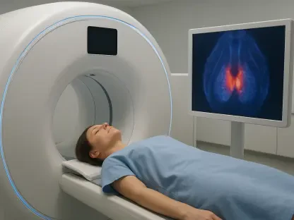
While molecular tests provide the “if” of a diagnosis, advanced imaging techniques have revolutionized the “where” and “how much” by providing detailed anatomical mapping without surgery. High-resolution transvaginal ultrasound, when performed by specially trained sonographers using standardized protocols, can now detect even small nodules of deep infiltrating disease. These technicians use specific maneuvers and bowel prep techniques to visualize adhesions and organ mobility in real-time, providing a dynamic view that a static MRI might miss. This specialized imaging allows for the creation of a “surgical map” before a patient ever enters the operating room, which significantly reduces operative time and improves the completeness of lesion excision. In many cases, a high-quality scan can provide enough evidence to skip diagnostic laparoscopy entirely and move straight to a therapeutic procedure if surgery is required. This precision ensures that patients receive the most appropriate level of care based on their specific anatomical involvement.
Future Implementation: Establishing New Standards of Care
The successful deployment of these non-invasive diagnostic tools throughout 2026 transformed the clinical landscape by finally prioritizing the lived experience of the patient over rigid surgical requirements. Medical professionals moved beyond the outdated “wait and see” philosophy, adopting a multi-modal approach that combined molecular screening with high-resolution imaging to deliver rapid results. For the first time, healthcare providers had the evidence needed to advocate for their patients’ needs with insurance companies and employers, citing objective biological data rather than subjective pain scores. To maintain this momentum, health systems prioritized the training of more sonographers in specialized endometriosis protocols and integrated microRNA testing into standard gynecological check-ups. Looking forward, the focus shifted toward using these non-invasive markers to tailor personalized medicine regimes, ensuring that every individual received treatment suited to their specific molecular profile. This evolution ensured that the era of the “invisible disease” was effectively ended, replaced by a new standard of evidence-based care.