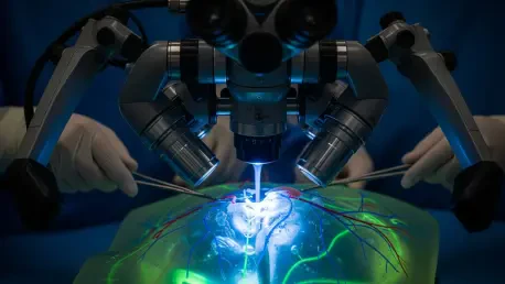

The ability of a surgeon to navigate the intricate landscape of the human body often rests on the clarity provided by near-infrared fluorescence imaging, a technology that has relied on Indocyanine Green for over sixty years. While this traditional dye has served as the gold standard for visualizing tumors and mapping lymphatic systems, it possesses a fundamental flaw that complicates modern medical procedures. When Indocyanine Green is subjected to the continuous laser light required for real-time visualization, it undergoes a process known as photobleaching, where the fluorescent signal rapidly loses its intensity. This degradation often forces medical teams to race against a fading visual guide, potentially compromising the accuracy of delicate dissections. As surgeries become increasingly complex and require longer durations of visibility, the need for a resilient imaging agent has become a critical priority for chemical engineers who seek to improve surgical outcomes.
Structural Resilience: The Science Behind Polymerized Dyes
To overcome the inherent instability of traditional fluorescence agents, researchers at the Korea Research Institute of Chemical Technology and Georgia Tech developed a novel substance known as KR-NIR-P. The core innovation lies in the transition from using isolated dye molecules to a sophisticated polymerized structure where the active components are integrated into a robust chemical backbone. This polymerization creates a physical shield that effectively isolates the dye from oxygen and other environmental factors that typically accelerate degradation. By anchoring the molecules within this stable architecture, the team successfully prevented the rapid chemical breakdown that ruins conventional alternatives under intense light. This approach represents a shift in strategy, moving away from temporary additives and toward a structural solution that maintains molecular integrity. The result is a more durable fluorescent signal that can withstand the rigors of modern surgical lighting.
The performance gap between KR-NIR-P and existing imaging standards is particularly evident when measuring the duration of the fluorescent signal under continuous laser exposure. In standard clinical scenarios, Indocyanine Green can lose more than half of its initial brightness in less than sixty seconds, leaving surgeons with a rapidly diminishing view of the surgical field. In contrast, the new polymerized dye demonstrated a remarkable ability to maintain its luminosity for extended periods, showing a four-fold improvement in overall stability during rigorous testing. This extended lifespan provides a significantly larger imaging window, ensuring that the visual guidance remains consistent and reliable from the beginning of a procedure to its final stages. By eliminating the anxiety associated with a fading signal, this technology allows surgical teams to focus entirely on the nuances of the operation. The reliability of this constant illumination is essential for high-stakes procedures.
Clinical Integration: Safety and Diagnostic Accuracy
Establishing a profile of biological safety is a mandatory prerequisite for any material destined for clinical use, and KR-NIR-P has undergone extensive testing to meet these requirements. Data collected from various cell line studies indicated that the dye is highly biocompatible, with over ninety percent of cells remaining healthy even when exposed to high concentrations of the polymer. The material does not trigger the biological markers associated with cell death, suggesting that it poses no greater risk than the current industry standards used in operating rooms today. This favorable safety profile is a critical step in the transition from research to human applications, providing the necessary evidence for regulatory approval. Beyond its safety, the dye demonstrates an impressive ability to interact with biological tissues without causing adverse reactions. This balance of performance and toxicity makes it an ideal candidate for next-generation intraoperative imaging tools.
In addition to its safety, the practical efficacy of KR-NIR-P has been validated through its performance in complex three-dimensional tumor models and animal studies focused on lymphatic mapping. The dye possesses a unique ability to penetrate deep into tumor tissues, providing high-contrast visuals that allow for the precise identification of malignant borders. This is particularly valuable in oncology, where the accurate mapping of the sentinel lymph nodes is vital for determining the extent of cancer metastasis. By gathering effectively within these nodes, the dye offers real-time diagnostic information that can influence the surgical strategy and the extent of tissue removal. The high-definition visibility provided by this material ensures that even small structures are clearly visible to the surgical team, reducing the likelihood of leaving cancerous cells behind. This performance confirms its potential as a tool for enhancing the precision of medical interventions.
Future Implementation: Preclinical Trials and Global Reach
The roadmap for the deployment of KR-NIR-P involves a series of detailed preclinical trials that will take place between 2026 and 2028 to monitor how the body processes the polymer. These studies are designed to observe the metabolic pathways and the eventual elimination of the material from the system, ensuring that no long-term accumulation occurs in vital organs. Supported by the Korean government and recognized in prominent scientific journals, this research highlights the intersection of chemical engineering and clinical medicine. The success of these upcoming trials is paramount for establishing the dye as a standard component of the surgical toolkit on a global scale. As medical facilities continue to upgrade their imaging capabilities, the demand for stable dyes will only increase. By providing a solution to the problem of photobleaching, this project sets a new benchmark for what is possible in the realm of fluorescent imaging and diagnostics.
The transition of KR-NIR-P into standard medical practice required hospital administrators to update imaging protocols to accommodate the longer window of visibility provided by the new polymer. Stakeholders recognized that training surgical staff on the subtle differences in fluorescence intensity helped maximize the benefits of the technology during complex procedures. The implementation of this dye encouraged researchers to explore similar polymerization techniques for other diagnostic markers, potentially revolutionizing how multi-spectral imaging is used in the operating room. Medical institutions that adopted this advancement early noted an improvement in surgical precision and a reduction in the time spent re-injecting traditional dyes. Ultimately, the successful deployment of this innovation proved that focusing on molecular stability was the most effective path toward long-term improvements in surgical navigation. Future engineering efforts focused on diversifying the color spectrum of these dyes.