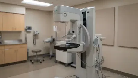

A prospective study of 415 women has demonstrated that Contrast-Enhanced Mammography effectively identifies hidden multifocality that traditional digital mammography frequently fails to detect. This breakthrough addresses a persistent challenge in breast oncology where determining the exact extent of a tumor determines the success of the initial surgery. For years, clinicians have struggled with the limitations of standard imaging, which often underestimates the spread of disease, leading to stressful and costly follow-up procedures. By integrating intravenous contrast agents with traditional mammographic techniques, researchers are finding that they can visualize blood flow to tumors, providing a clearer map of the malignancy. This Swedish trial, conducted at Skane University Hospital, represents a significant step forward in validating this technology for early-stage breast cancer. The focus remains on whether this enhanced clarity translates into better surgical outcomes or if the diagnostic precision is currently ahead of clinical practice.
Comparative Diagnostic Precision
Evaluating Study Protocols and Demographic Data
The study was structured as a rigorous prospective randomized controlled trial to ensure that the findings were both statistically significant and clinically relevant. It focused on a cohort of 415 women with a median age of 66, all of whom had been diagnosed with early-stage breast cancer and were scheduled for primary surgery. To facilitate a clear comparison, the participants were split into two distinct groups. The experimental group, consisting of 202 women, received preoperative Contrast-Enhanced Mammography in addition to standard imaging. The control group received the current standard of care, which typically includes digital mammography, digital breast tomosynthesis, and handheld ultrasound. This division allowed the researchers to isolate the specific impact of CEM on the preoperative staging process. By comparing these two pathways, the trial sought to determine if the additional data provided by contrast imaging would lead to more precise surgical plans and a reduction in the need for second operations.
Measuring Accuracy and Malignant Extent
One of the most compelling findings from the research was the significant difference in how accurately each imaging modality estimated the actual extent of the cancer. When the preoperative images were compared to the postoperative histopathology, CEM emerged as the most precise tool available to the surgical team. It showed a measurement bias of only 3.5 millimeters, meaning its estimates were remarkably close to the actual size of the tumor as confirmed under a microscope. In comparison, digital mammography and digital breast tomosynthesis displayed a wider bias of 4.8 millimeters, while handheld ultrasound was the least accurate, trailing with a significant bias of 10.7 millimeters. This accuracy was not just a general trend but was consistent across various patient demographics, including those with high breast density. This is particularly important because dense tissue often obscures tumors on traditional mammograms, yet CEM appears to bypass this hurdle by focusing on vascular patterns rather than tissue density.
Clinical Application and Systemic Challenges
Surgical Modifications and Treatment Decisions
The enhanced diagnostic clarity of CEM directly influenced the surgical management of approximately 8 percent of the patients in the experimental group. In 17 specific cases, the information provided by the contrast scan led the oncology team to alter the planned surgical approach. For 16 of these individuals, the changes were later confirmed to be correct through histopathologic analysis of the removed tissue. Most of these adjustments involved a shift from a planned partial mastectomy to a full mastectomy because the CEM images revealed additional tumor sites that were invisible on standard scans. For instance, in one notable case, initial imaging suggested a single 16-millimeter lesion, but CEM identified a complex network of at least six additional spots that extended the total malignant area to 80 millimeters. Without this advanced imaging, the surgeon likely would have performed an inadequate procedure, necessitating a second, more invasive operation later. This highlights the technology’s role in ensuring oncologic safety from the start.
Addressing Paradoxical Outcomes and Institutional Barriers
Despite the undeniable precision of CEM in measuring tumors, the trial revealed a surprising paradox: the overall rates of mastectomy and reoperation did not show a statistically significant decrease compared to the control group. The mastectomy rate stood at 17 percent for the CEM group versus 18 percent for the control, while reoperation rates were 7.9 percent and 10 percent, respectively. Researchers believe this discrepancy stems from deep-seated institutional preferences and a reliance on traditional surgical thresholds. At several study sites, medical teams maintained a strong historical preference for breast-conserving therapy and were hesitant to pivot to more aggressive procedures based on new technology alone. This suggests that the successful integration of CEM depends as much on cultural shifts within the surgical community as it does on the technical merits of the imaging itself. If surgeons do not fully trust the additional findings or lack standardized protocols for acting on them, the clinical benefits may remain underutilized.
Concluding Synthesis and Strategic Recommendations
In conclusion, the trial established that Contrast-Enhanced Mammography offered a more precise evaluation of tumor size than any other current imaging method. The researchers found that while the technology successfully identified occult lesions and guided more accurate surgical planning for a specific subset of patients, its impact on total population statistics remained limited by existing clinical habits. The study noted that future implementations should focus on expanding access to CEM-guided biopsy equipment to verify findings without reverting to older modalities. Furthermore, the medical community moved toward developing standardized multidisciplinary guidelines that specifically addressed how to manage multifocality detected only by CEM. The findings suggested that for high-risk patients or those with complex presentations, CEM proved to be an indispensable asset for avoiding surgical failure. As surgeons gained more confidence in these digital insights, the potential for CEM to refine breast cancer staging and personalize surgical care became increasingly clear.