
Current clinical workflows for diagnosing hip impingement often rely on CT scans that expose patients to ionizing radiation or expensive, time-consuming MRI procedures. This persistent diagnostic hurdle has driven a multidisciplinary team of biomedical engineers and surgeons at Dalhousie University to develop a radiation-free alternative that matches the precision of traditional imaging. Led by researchers Andrew D. Michels and orthopedic surgeon Ivan Wong, the team introduced a freehand 3D ultrasound system designed to visualize the complex geometry of the hip joint. Their primary target is femoroacetabular impingement syndrome (FAIS), a common musculoskeletal disorder where abnormal contact occurs between the femoral head and the hip socket. This friction is typically caused by a “cam deformity,” an excess protrusion of bone that strikes the rim of the socket during movement. Because FAIS predominantly affects young, active populations, the cumulative risk of ionizing radiation from repeated CT scans is a significant clinical concern. This new ultrasound method offers a breakthrough by providing the necessary three-dimensional detail for surgical planning without the biological risks or the high financial costs associated with existing modalities.
Advanced AI and Deep Learning Integration
To create a comprehensive three-dimensional map from what is traditionally a two-dimensional imaging tool, the researchers utilized a freehand ultrasound approach combined with sophisticated tracking technology. In this setup, a clinician sweeps a standard ultrasound probe over the patient’s hip while an optical tracking system continuously records the exact position and orientation of the probe in space. Every 2D “slice” captured by the ultrasound is then tagged with its spatial coordinates, allowing the software to assemble thousands of individual frames into a cohesive 3D volume. However, the true technical bottleneck in this process has historically been segmentation, which is the difficult task of distinguishing the bright line of the bone from the surrounding soft tissue, shadows, and artifacts. Manually tracing these bone surfaces across thousands of frames would be an impossible task for a clinician in a fast-paced medical environment, necessitating an automated solution that can process data in real-time.
The research team solved this processing challenge by implementing a specialized deep learning architecture known as a Feature Pyramid Network (FPN). This convolutional neural network was specifically trained to detect and isolate bone surfaces within the ultrasound images with high reliability. To ensure the system was effective despite having a relatively small amount of specialized hip data, the engineers employed a technique called transfer learning. They began with a model already familiar with general bone structures from larger, public datasets and then fine-tuned the AI using a specific set of manually segmented hip images. This approach allowed the system to achieve exceptional accuracy in identifying the cortical surface of the femur without requiring tens of thousands of new labeled images. This integration of artificial intelligence transforms a standard ultrasound scan into a high-fidelity diagnostic tool, effectively removing the human labor component that previously hindered the clinical adoption of 3D ultrasound for bone modeling.
Hardware Efficiency and Digital Reconstruction
A core advantage of the Dalhousie system is its commitment to using affordable, off-the-shelf hardware components rather than proprietary, high-cost equipment. The system pairs a portable point-of-care ultrasound probe with a standard optical tracker, a configuration that is significantly more economical than the multi-million dollar infrastructure required for CT or MRI suites. This hardware efficiency suggests that the technology could be readily deployed in smaller orthopedic clinics, rural hospitals, or specialized sports medicine facilities where high-end imaging may be unavailable. By lowering the financial and logistical barriers to advanced imaging, the team is paving the way for a more decentralized model of orthopedic care. Patients who previously had to wait weeks for an MRI or travel to major medical centers for a CT scan could potentially receive a full 3D diagnostic workup during their initial consultation with a specialist.
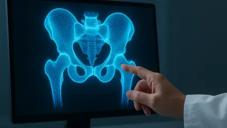
Once the AI-driven software identifies the bone boundaries from the tracked ultrasound frames, deterministic algorithms convert these data points into a point cloud representing the surface of the femoral head and neck junction. The software then processes this cloud into a “watertight surface mesh,” which is essentially a smooth, continuous digital twin of the patient’s actual bone structure. This digital reconstruction provides a level of spatial awareness that 2D X-rays cannot match, as it allows surgeons to rotate the model, zoom in on specific anatomical features, and inspect the joint from angles that are physically impossible to view using standard equipment. This capability is vital for quantifying the exact size and location of a cam deformity. The resulting digital twin serves as a precise roadmap for the surgeon, ensuring that any subsequent arthroscopic procedure is as targeted and minimally invasive as possible, ultimately leading to better postoperative outcomes and faster recovery times.
Validation Against Clinical Gold Standards
To prove that this low-cost method could stand up to established diagnostic tools, the researchers conducted a rigorous validation study comparing their ultrasound models to CT scans, which are considered the gold standard for bone geometry. The team tested the system on both cadaveric hip specimens and living subjects to ensure the results were repeatable and consistent across different operators. The findings were highly encouraging, showing a geometric agreement between the 3D ultrasound meshes and CT scans of 0.4 to 0.6 millimeters. In the context of orthopedic surgery for FAIS, bone deformities are typically considered clinically significant if they exceed two millimeters. Because the ultrasound system’s margin of error is less than one millimeter, it proved its ability to reliably detect even the most subtle structural irregularities that might lead to joint degradation or chronic pain if left unaddressed.
Beyond general geometric accuracy, the system was evaluated using the “alpha angle,” which is the primary metric used by surgeons to determine the severity of a hip deformity and the need for surgery. An alpha angle exceeding a certain threshold indicates a loss of the normal spherical shape of the femoral head. The study revealed that the measurements derived from the 3D ultrasound models agreed with CT-based measurements to within 0.6 degrees. Since a variance of five degrees is generally the threshold for clinical significance, the system’s error rate is nearly ten times smaller than what is required for a confident diagnosis. These results were further bolstered by in vivo tests on healthy volunteers, which demonstrated that the workflow remained effective when scanning through skin and muscle tissue in a real-world setting. This level of precision confirms that the system is not merely a laboratory curiosity but a robust clinical tool ready for further development.
Future Applications in Orthopedic Care
The success of this automated bone mapping technique has implications that extend far beyond the narrow scope of hip impingement. By demonstrating that 3D ultrasound can provide CT-like detail for bony surfaces, the Dalhousie team has provided a blueprint for various other orthopedic applications. For instance, the technology could be adapted for monitoring the progression of osteoarthritis over several years, from 2026 to 2030, without subjecting patients to repeated doses of ionizing radiation. It also shows great promise for evaluating the alignment and healing of complex fractures in real-time at the point of care. Perhaps most significantly, the system could revolutionize pediatric orthopedics. In cases like hip dysplasia, where children require frequent monitoring of their developing joints, the ability to avoid radiation exposure is a paramount safety advantage that could prevent long-term health complications related to medical imaging.
The transition from a laboratory prototype to a standard clinical tool will require further expansion of the current research. While the validation results were impressive, the researchers recognized that the initial study involved a relatively small group of participants. Larger, multi-center clinical trials are now the necessary next step to ensure the system performs consistently across diverse patient populations with varying body types, ages, and anatomical complexities. There is also a practical need to refine the optical tracking interface to make it even more user-friendly for clinicians who may not have specialized engineering training. Despite these remaining hurdles, the development of the freehand 3D ultrasound system represented a major victory for diagnostic safety and efficiency. The project effectively mapped a new standard of care, ensuring that precise orthopedic diagnostics became more accessible, less expensive, and significantly safer for patients across the globe.